

152
can change under several stressful conditions, such
as heat exposure, handling stress, hyperoxia, oxidized oils administration, and heavy metal intake
(Parihar and Dubey 1995 ; Nakano et al. 1999a ; Ali
et al. 2004 ; Rau et al. 2004 ; Lushchak et al. 2005a, b ;
Martínez-Álvarez et al. 2005 ; Olsen et al. 2005 ;
Heise et al. 2006 ; Lesser 2006 ; Valavanidis et al.
2006 ; Bagnyukova et al. 2007 ).
The major nonprotein cellular thiol, reduced
GSH, is a tripeptide (Glu-Cys-Gly) with reducing and nucleophilic properties that is one of the
major regulators of the intracellular redox state
(Niki 1988 ; Nakano and Takeuchi 1997 ; Arrigo
1999 ; Sies 1999 ; Lesser 2006 ; Valavanidis et al.
2006 ). GSH can act as a chain breaker of free
radical reaction and is the substrate for glutathione peroxidase, an enzyme that scavenges reactive oxygen species (ROS) and LPO generated
within cells. The plasma GSH levels observed in
this study were similar to those in the livers of
fi sh that were administered with an oxidant,
such as t-butyl hydroperoxide, after heat exposure (Ploch et al. 1999 ; Ali et al. 2004 ; Lushchak
and Bagnyukova 2006a, b ; Heise et al. 2006 ;
Valavanidis et al. 2006 ; Bagnyukova et al.
2007 ). At the initial post-heat stress stage, GSH
may be consumed to eliminate ROS generated
in blood.
Plasma SOD activity in heat-shocked fi sh
showed a transient increase at 17.5 h post-stress
in this study. Antioxidative enzymes, such as
SOD, glutathione peroxidase, and catalase, can
scavenge radicals and contribute to the body’s
antioxidative defenses. In particular, SODs are
considered to have key roles in the fi rst line of
the enzymatic antioxidative defense system
against oxidative injuries, as they catalyze the
removal of oxygen radical, superoxide (Asada
1988 ; Oyanagui 1989 ; Nakano and Takeuchi
1997 ; Taniguchi and Endo 2000 ; Zelko et al.
2002 ; Martínez-Álvarez et al. 2005 ; Lesser
2006 ; Lushchak 2011 ). Hence, increased SOD
expression may neutralize the harmful effects of
superoxides in tissues. The changes in the
expression of antioxidative enzymes, such as
SOD, in fi sh have been observed with regard to
stress (Poly 1997 ; Pörtner 2002 ; MartínezÁlvarez et al. 2005 ; Valavanidis et al. 2006 ;
Craig et al. 2007 ; Lushchak 2011 ).
Heat exposure and enhanced oxygen consumption are considered to promote ROS generation in the tissue. The resulting ROS attack
almost all cell components (Asada 1988 ; Nakano
and Takeuchi 1997 ; Beckman and Ames 1998 ;
Droge 2002 ; Lesser 2006 ; Valavanidis et al.
2006 ; Lushchak 2011 ). ROS production in cell,
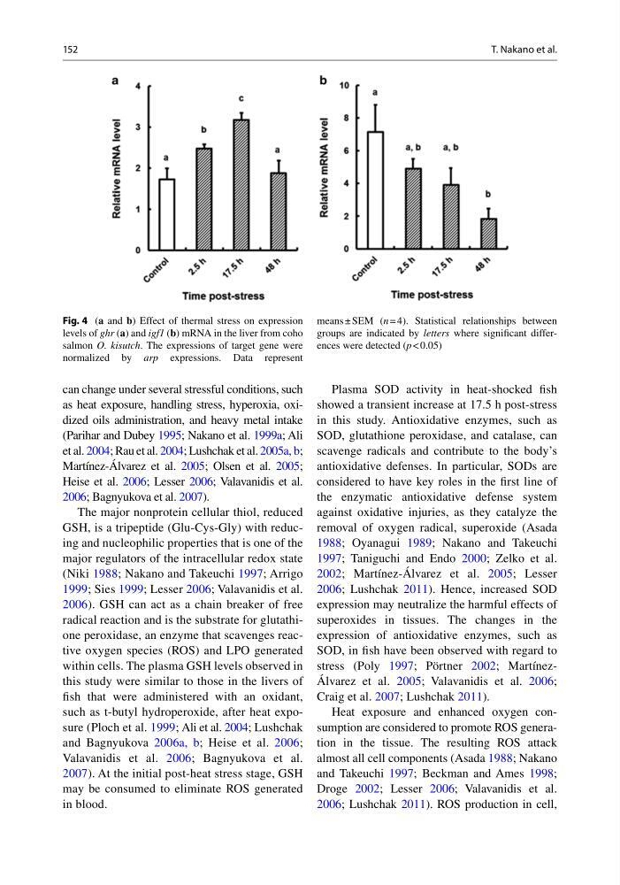
Fig. 4 ( a and b ) Effect of thermal stress on expression
levels of ghr ( a ) and igf1 ( b ) mRNA in the liver from coho
salmon O. kisutch . The expressions of target gene were
normalized by arp expressions. Data represent
means ± SEM ( n = 4). Statistical relationships between
groups are indicated by letters where signifi cant differences were detected ( p < 0.05)
T. Nakano et al.
can change under several stressful conditions, such
as heat exposure, handling stress, hyperoxia, oxidized oils administration, and heavy metal intake
(Parihar and Dubey 1995 ; Nakano et al. 1999a ; Ali
et al. 2004 ; Rau et al. 2004 ; Lushchak et al. 2005a, b ;
Martínez-Álvarez et al. 2005 ; Olsen et al. 2005 ;
Heise et al. 2006 ; Lesser 2006 ; Valavanidis et al.
2006 ; Bagnyukova et al. 2007 ).
The major nonprotein cellular thiol, reduced
GSH, is a tripeptide (Glu-Cys-Gly) with reducing and nucleophilic properties that is one of the
major regulators of the intracellular redox state
(Niki 1988 ; Nakano and Takeuchi 1997 ; Arrigo
1999 ; Sies 1999 ; Lesser 2006 ; Valavanidis et al.
2006 ). GSH can act as a chain breaker of free
radical reaction and is the substrate for glutathione peroxidase, an enzyme that scavenges reactive oxygen species (ROS) and LPO generated
within cells. The plasma GSH levels observed in
this study were similar to those in the livers of
fi sh that were administered with an oxidant,
such as t-butyl hydroperoxide, after heat exposure (Ploch et al. 1999 ; Ali et al. 2004 ; Lushchak
and Bagnyukova 2006a, b ; Heise et al. 2006 ;
Valavanidis et al. 2006 ; Bagnyukova et al.
2007 ). At the initial post-heat stress stage, GSH
may be consumed to eliminate ROS generated
in blood.
Plasma SOD activity in heat-shocked fi sh
showed a transient increase at 17.5 h post-stress
in this study. Antioxidative enzymes, such as
SOD, glutathione peroxidase, and catalase, can
scavenge radicals and contribute to the body’s
antioxidative defenses. In particular, SODs are
considered to have key roles in the fi rst line of
the enzymatic antioxidative defense system
against oxidative injuries, as they catalyze the
removal of oxygen radical, superoxide (Asada
1988 ; Oyanagui 1989 ; Nakano and Takeuchi
1997 ; Taniguchi and Endo 2000 ; Zelko et al.
2002 ; Martínez-Álvarez et al. 2005 ; Lesser
2006 ; Lushchak 2011 ). Hence, increased SOD
expression may neutralize the harmful effects of
superoxides in tissues. The changes in the
expression of antioxidative enzymes, such as
SOD, in fi sh have been observed with regard to
stress (Poly 1997 ; Pörtner 2002 ; MartínezÁlvarez et al. 2005 ; Valavanidis et al. 2006 ;
Craig et al. 2007 ; Lushchak 2011 ).
Heat exposure and enhanced oxygen consumption are considered to promote ROS generation in the tissue. The resulting ROS attack
almost all cell components (Asada 1988 ; Nakano
and Takeuchi 1997 ; Beckman and Ames 1998 ;
Droge 2002 ; Lesser 2006 ; Valavanidis et al.
2006 ; Lushchak 2011 ). ROS production in cell,
Fig. 4 ( a and b ) Effect of thermal stress on expression
levels of ghr ( a ) and igf1 ( b ) mRNA in the liver from coho
salmon O. kisutch . The expressions of target gene were
normalized by arp expressions. Data represent
means ± SEM ( n = 4). Statistical relationships between
groups are indicated by letters where signifi cant differences were detected ( p < 0.05)
T. Nakano et al.