

g Applications in Genetics
Single-Cell
Experiments
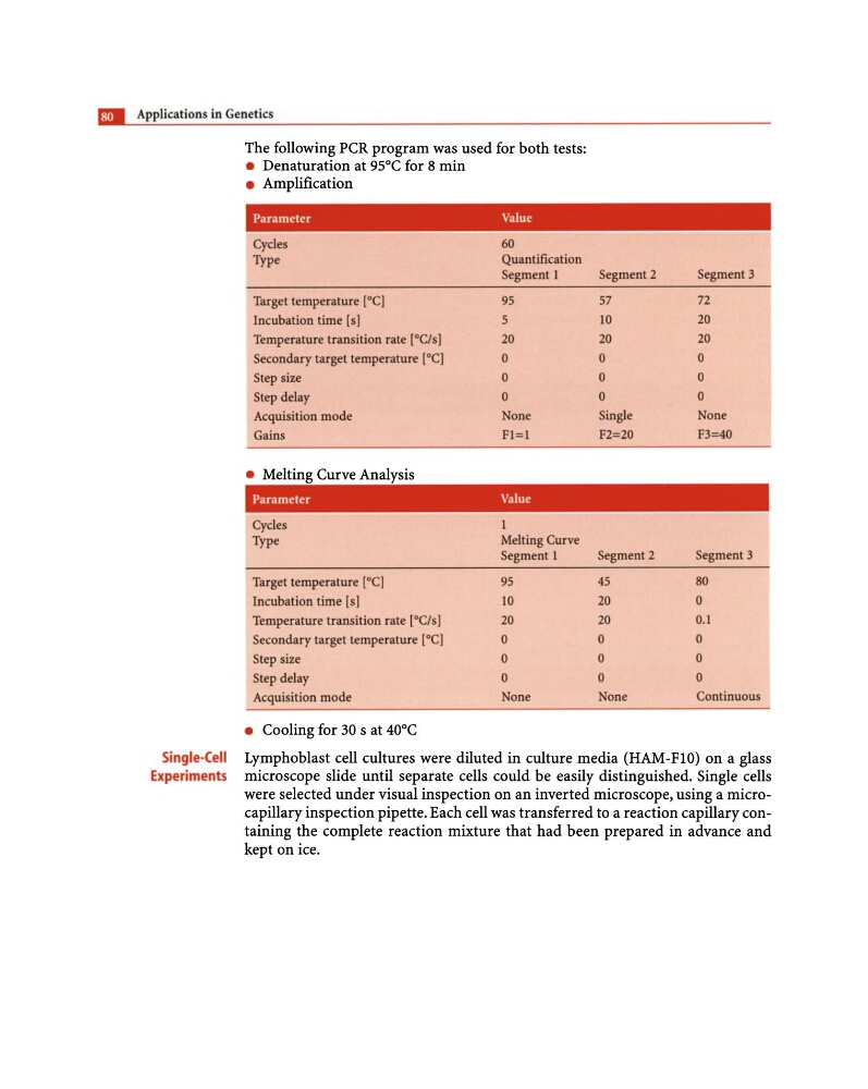
The following PCR program was used for both tests:
• Denaturation at 95°C for 8 min
• Amplification
Parameter
Va lue
Cycles
60
Type
Quantification
Segment 1
Segment 2
Segment 3
Target temperature [0C]
95
57
72
Incubation time [s]
5
10
20
Temperature transition rate rOC/sI
20
20
20
Secondary target temperature [0C]
0
0
0
Step size
0
0
0
Step delay
0
0
0
Acquisition mode
None
Single
None
Gains
Fl=l
F2=20
F3=40
• Melting Curve Analysis
Parameter
Va llie
Cycles
Type
Melting Curve
Segment 1
Segment 2
Segment 3
Target temperature [0C)
95
45
80
Incubation time [s]
10
20
0
Temperature transition rate [OC/s J
20
20
0.1
Secondary target temperature [0C]
0
0
0
Step size
0
0
0
Step delay
0
0
0
Acquisition mode
None
None
Continuous
• Cooling for 30 s at 40°C
Lymphoblast cell cultures were diluted in culture media (HAM-FlO) on a glass
microscope slide until separate cells could be easily distinguished. Single cells
were selected under visual inspection on an inverted microscope, using a microcapillary inspection pipette. Each cell was transferred to a reaction capillary containing the complete reaction mixture that had been prepared in advance and
kept on ice.
Single-Cell
Experiments
The following PCR program was used for both tests:
• Denaturation at 95°C for 8 min
• Amplification
Parameter
Va lue
Cycles
60
Type
Quantification
Segment 1
Segment 2
Segment 3
Target temperature [0C]
95
57
72
Incubation time [s]
5
10
20
Temperature transition rate rOC/sI
20
20
20
Secondary target temperature [0C]
0
0
0
Step size
0
0
0
Step delay
0
0
0
Acquisition mode
None
Single
None
Gains
Fl=l
F2=20
F3=40
• Melting Curve Analysis
Parameter
Va llie
Cycles
Type
Melting Curve
Segment 1
Segment 2
Segment 3
Target temperature [0C)
95
45
80
Incubation time [s]
10
20
0
Temperature transition rate [OC/s J
20
20
0.1
Secondary target temperature [0C]
0
0
0
Step size
0
0
0
Step delay
0
0
0
Acquisition mode
None
None
Continuous
• Cooling for 30 s at 40°C
Lymphoblast cell cultures were diluted in culture media (HAM-FlO) on a glass
microscope slide until separate cells could be easily distinguished. Single cells
were selected under visual inspection on an inverted microscope, using a microcapillary inspection pipette. Each cell was transferred to a reaction capillary containing the complete reaction mixture that had been prepared in advance and
kept on ice.