

Genotyping by Guanosine-Dependent Quenching of Single-Labeled Fluorescein Probes IIjJ
50 ~~----------------------------------------,
Q)
() 45
c:
Q)
()
CJ)
Q)
~
o
:::J
LL 40
0.5
r
1:)
0
::::::...
~
LL
'-"
1:)
I -0.5
A
-1
B
40
- " - Wild Type
-
Heterozygous
- - - Homozygous Mutant
........ No D NA
... . . '.
50
60
70
Temperature (O C)
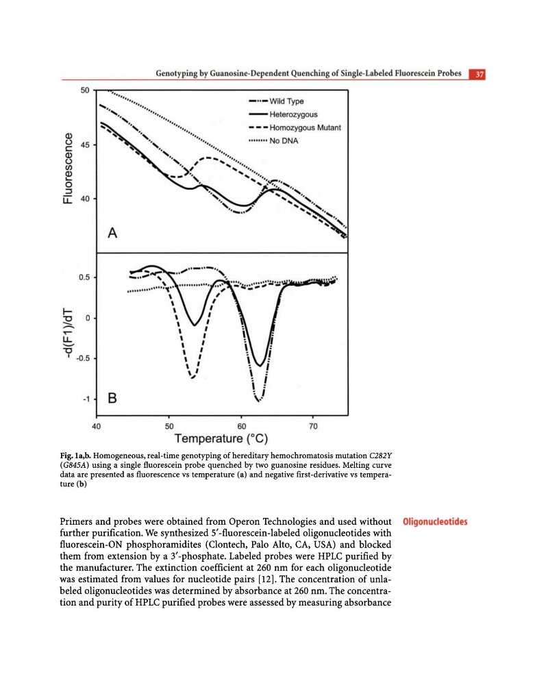
Fig. la,b. Homogeneous, real-time genotyping of hereditary hemochromatosis mutation C282Y
(G845A) using a single fluorescein probe quenched by two guanosine residues. Melting curve
data are presented as fluorescence vs temperature (a) and negative first-derivative vs temperature (b)
Primers and probes were obtained from Operon Technologies and used without Oligonucleotides
further purification. We synthesized 5'-fluorescein -labeled oligonucleotides with
fluorescein-ON phosphoramidites (Clontech, Palo Alto, CA, USA) and blocked
them from extension by a 3'-phosphate. Labeled probes were HPLC purified by
the manufacturer. The extinction coefficient at 260 nm for each oligonucleotide
was estimated from values for nucleotide pairs [12]. The concentration of unlabeled oligonucleotides was determined by absorbance at 260 nm. The concentration and purity of HPLC purified probes were assessed by measuring absorbance
50 ~~----------------------------------------,
Q)
() 45
c:
Q)
()
CJ)
Q)
~
o
:::J
LL 40
0.5
r
1:)
0
::::::...
~
LL
'-"
1:)
I -0.5
A
-1
B
40
- " - Wild Type
-
Heterozygous
- - - Homozygous Mutant
........ No D NA
... . . '.
50
60
70
Temperature (O C)
Fig. la,b. Homogeneous, real-time genotyping of hereditary hemochromatosis mutation C282Y
(G845A) using a single fluorescein probe quenched by two guanosine residues. Melting curve
data are presented as fluorescence vs temperature (a) and negative first-derivative vs temperature (b)
Primers and probes were obtained from Operon Technologies and used without Oligonucleotides
further purification. We synthesized 5'-fluorescein -labeled oligonucleotides with
fluorescein-ON phosphoramidites (Clontech, Palo Alto, CA, USA) and blocked
them from extension by a 3'-phosphate. Labeled probes were HPLC purified by
the manufacturer. The extinction coefficient at 260 nm for each oligonucleotide
was estimated from values for nucleotide pairs [12]. The concentration of unlabeled oligonucleotides was determined by absorbance at 260 nm. The concentration and purity of HPLC purified probes were assessed by measuring absorbance