

Rapid Quantitative Detection of Free Cancer Cells in the Peritoneal Cavity of Gastric Cancer Patients 1m
prove the integrity of the isolated RNA, a real-time RT-PCR analysis for glyceraldehyde-3-phosphate dehydrogenase (GAPDH) was also carried out using
primers and hybridization probes [5].
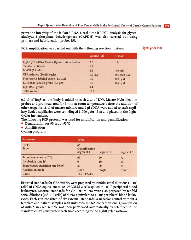
PCR amplification was carried out with the following reaction mixture:
Volume 1l1iJ
[FinalJ
LightCycier-DNA Master Hybridization Probes
2.0
IX
TaqStart antibody
0.2
MgCI2 (25 mM)
2.4
4.0mM
CEA primers (10 flM each)
1.0+1.0
0.5 each flM
Fluorescein-labeled probe (4.0 flM)
1.0
0.20 flM
LCRed640-labeled probe (8.0 flM)
1.0
DAD flM
H20 (PCR grade)
9.4
Total volume
18.0
0.2 fll of TaqStart antibody is added to each 2 fll of DNA Master Hybridization
probes and pre-incubated for 5 min at room temperature before the addition of
other reagents. 18 f.Ll of master mixture and 2 f.Ll cDNA were added to each capillary. Sealed capillaries were centrifuged (1000 g for 15 s) and placed in the LightCycler instrument.
The following PCR protocol was used for amplification and quantification:
• Denaturation for 90 sec at 95°C
• Amplification
Cycling program:
Parameter
Value
Cycles
45
Type
Quantification
Segment 1
Target temperature [0C]
95
lncubation time [s)
0
Temperature transition rate lOC/s]
20
Acquisition mode
None
Gains
Fl=l; F2=15
Segment 2
Segment 3
50
72
10
10
20
20
Single
None
External standards for CEA mRNA were prepared by tenfold serial dilutions (1-10 5
cells) of cDNA equivalent to lxl0 6 COLM-2 cells spiked to lx10 7 peripheral blood
leukocytes. External standards for GAPDH mRNA were also prepared by tenfold
serial dilutions (10 2 -10 7 cells) of cDNA equivalent to 1 x 1 0 7 peripheral blood leukocytes. Each run consisted of six external standards, a negative control without a
template and patient samples with unknown mRNA concentrations. Quantitation
of mRNA in each sample was then performed automatically by reference to the
standard curve constructed each time according to the LightCycler software.
LightCyder PCR
prove the integrity of the isolated RNA, a real-time RT-PCR analysis for glyceraldehyde-3-phosphate dehydrogenase (GAPDH) was also carried out using
primers and hybridization probes [5].
PCR amplification was carried out with the following reaction mixture:
Volume 1l1iJ
[FinalJ
LightCycier-DNA Master Hybridization Probes
2.0
IX
TaqStart antibody
0.2
MgCI2 (25 mM)
2.4
4.0mM
CEA primers (10 flM each)
1.0+1.0
0.5 each flM
Fluorescein-labeled probe (4.0 flM)
1.0
0.20 flM
LCRed640-labeled probe (8.0 flM)
1.0
DAD flM
H20 (PCR grade)
9.4
Total volume
18.0
0.2 fll of TaqStart antibody is added to each 2 fll of DNA Master Hybridization
probes and pre-incubated for 5 min at room temperature before the addition of
other reagents. 18 f.Ll of master mixture and 2 f.Ll cDNA were added to each capillary. Sealed capillaries were centrifuged (1000 g for 15 s) and placed in the LightCycler instrument.
The following PCR protocol was used for amplification and quantification:
• Denaturation for 90 sec at 95°C
• Amplification
Cycling program:
Parameter
Value
Cycles
45
Type
Quantification
Segment 1
Target temperature [0C]
95
lncubation time [s)
0
Temperature transition rate lOC/s]
20
Acquisition mode
None
Gains
Fl=l; F2=15
Segment 2
Segment 3
50
72
10
10
20
20
Single
None
External standards for CEA mRNA were prepared by tenfold serial dilutions (1-10 5
cells) of cDNA equivalent to lxl0 6 COLM-2 cells spiked to lx10 7 peripheral blood
leukocytes. External standards for GAPDH mRNA were also prepared by tenfold
serial dilutions (10 2 -10 7 cells) of cDNA equivalent to 1 x 1 0 7 peripheral blood leukocytes. Each run consisted of six external standards, a negative control without a
template and patient samples with unknown mRNA concentrations. Quantitation
of mRNA in each sample was then performed automatically by reference to the
standard curve constructed each time according to the LightCycler software.
LightCyder PCR