

12
c.c. Parrish
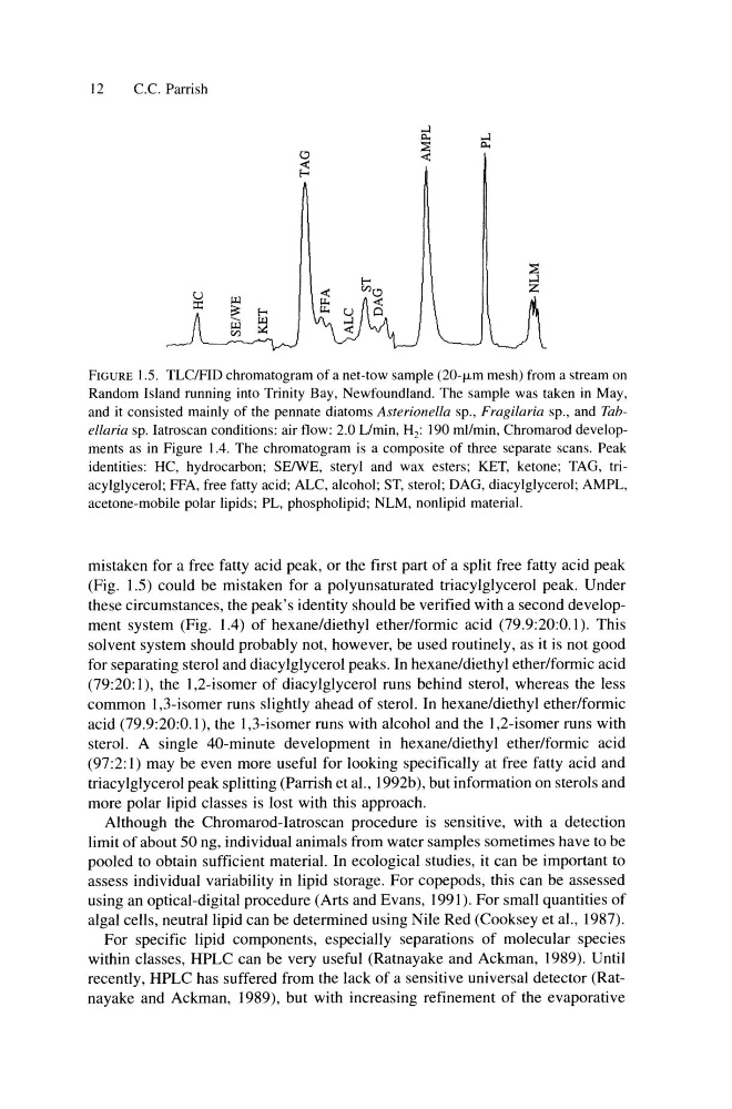
FIGURE 1.5. TLC/FID chromatogram of a net-tow sample (20-f.Lm mesh) from a stream on
Random Island running into Trinity Bay, Newfoundland. The sample was taken in May,
and it consisted mainly of the pennate diatoms Asterionella sp., Fragilaria sp., and Tabella ria sp. Iatroscan conditions: air flow: 2.0 Llmin, H 2 : 190 mllmin, Chromarod developments as in Figure 1.4. The chromatogram is a composite of three separate scans. Peak
identities: HC, hydrocarbon; SEIWE, steryl and wax esters; KET, ketone; TAG, triacylglycerol; FFA, free fatty acid; ALC, alcohol; ST, sterol; DAG, diacylglycerol; AMPL,
acetone-mobile polar lipids; PL, phospholipid; NLM, nonlipid material.
mistaken for a free fatty acid peak, or the first part of a split free fatty acid peak
(Fig. 1.5) could be mistaken for a polyunsaturated triacylglycerol peak. Under
these circumstances, the peak's identity should be verified with a second development system (Fig. 1.4) of hexane/diethyl ether/formic acid (79.9:20:0.1). This
solvent system should probably not, however, be used routinely, as it is not good
for separating sterol and diacylglycerol peaks. In hexane/diethyl ether/formic acid
(79:20:1), the 1,2-isomer of diacylglycerol runs behind sterol, whereas the less
common 1 ,3-isomer runs slightly ahead of sterol. In hexane/diethyl ether/formic
acid (79.9:20:0.1), the 1,3-isomer runs with alcohol and the I ,2-isomer runs with
sterol. A single 40-minute development in hexane/diethyl ether/formic acid
(97 :2: I) may be even more useful for looking specifically at free fatty acid and
triacylglycerol peak splitting (Parrish et aI., 1 992b), but information on sterols and
more polar lipid classes is lost with this approach.
Although the Chromarod-Iatroscan procedure is sensitive, with a detection
limit of about 50 ng, individual animals from water samples sometimes have to be
pooled to obtain sufficient material. In ecological studies, it can be important to
assess individual variability in lipid storage. For copepods, this can be assessed
using an optical-digital procedure (Arts and Evans, 1991). For small quantities of
algal cells, neutral lipid can be determined using Nile Red (Cooksey et aI., 1987).
For specific lipid components, especially separations of molecular species
within classes, HPLC can be very useful (Ratnayake and Ackman, 1989). Until
recently, HPLC has suffered from the lack of a sensitive universal detector (Ratnayake and Ackman, 1989), but with increasing refinement of the evaporative
c.c. Parrish
FIGURE 1.5. TLC/FID chromatogram of a net-tow sample (20-f.Lm mesh) from a stream on
Random Island running into Trinity Bay, Newfoundland. The sample was taken in May,
and it consisted mainly of the pennate diatoms Asterionella sp., Fragilaria sp., and Tabella ria sp. Iatroscan conditions: air flow: 2.0 Llmin, H 2 : 190 mllmin, Chromarod developments as in Figure 1.4. The chromatogram is a composite of three separate scans. Peak
identities: HC, hydrocarbon; SEIWE, steryl and wax esters; KET, ketone; TAG, triacylglycerol; FFA, free fatty acid; ALC, alcohol; ST, sterol; DAG, diacylglycerol; AMPL,
acetone-mobile polar lipids; PL, phospholipid; NLM, nonlipid material.
mistaken for a free fatty acid peak, or the first part of a split free fatty acid peak
(Fig. 1.5) could be mistaken for a polyunsaturated triacylglycerol peak. Under
these circumstances, the peak's identity should be verified with a second development system (Fig. 1.4) of hexane/diethyl ether/formic acid (79.9:20:0.1). This
solvent system should probably not, however, be used routinely, as it is not good
for separating sterol and diacylglycerol peaks. In hexane/diethyl ether/formic acid
(79:20:1), the 1,2-isomer of diacylglycerol runs behind sterol, whereas the less
common 1 ,3-isomer runs slightly ahead of sterol. In hexane/diethyl ether/formic
acid (79.9:20:0.1), the 1,3-isomer runs with alcohol and the I ,2-isomer runs with
sterol. A single 40-minute development in hexane/diethyl ether/formic acid
(97 :2: I) may be even more useful for looking specifically at free fatty acid and
triacylglycerol peak splitting (Parrish et aI., 1 992b), but information on sterols and
more polar lipid classes is lost with this approach.
Although the Chromarod-Iatroscan procedure is sensitive, with a detection
limit of about 50 ng, individual animals from water samples sometimes have to be
pooled to obtain sufficient material. In ecological studies, it can be important to
assess individual variability in lipid storage. For copepods, this can be assessed
using an optical-digital procedure (Arts and Evans, 1991). For small quantities of
algal cells, neutral lipid can be determined using Nile Red (Cooksey et aI., 1987).
For specific lipid components, especially separations of molecular species
within classes, HPLC can be very useful (Ratnayake and Ackman, 1989). Until
recently, HPLC has suffered from the lack of a sensitive universal detector (Ratnayake and Ackman, 1989), but with increasing refinement of the evaporative