

1. Lipid Determination in Aquatic Samples
11
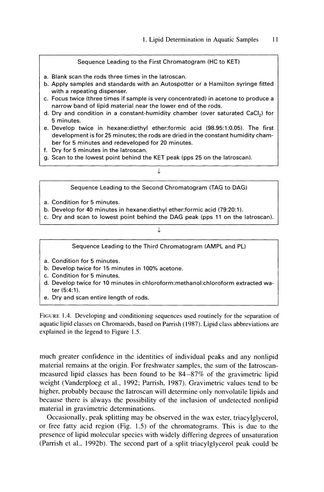
Sequence Leading to the First Chromatogram (HC to KET)
a. Blank scan the rods three times in the latroscan.
b. Apply samples and standards with an Autospotter or a Hamilton syringe fitted
with a repeating dispenser.
c. Focus twice (three times if sample is very concentrated) in acetone to produce a
narrow band of lipid material near the lower end of the rods.
d. Dry and condition in a constant-humidity chamber (over saturated CaCI 2 ) for
5 minutes.
e. Develop twice in hexane:diethyl ether:formic acid (98.95:1:0.05). The first
development is for 25 minutes; the rods are dried in the constant humidity chamber for 5 minutes and redeveloped for 20 minutes.
f. Dry for 5 minutes in the latroscan.
g. Scan to the lowest point behind the KET peak (pps 25 on the latroscan).
Sequence Leading to the Second Chromatogram (TAG to DAG)
a. Condition for 5 minutes.
b. Develop for 40 minutes in hexane:diethyl ether:formic acid (79:20:1).
c. Dry and scan to lowest point behind the DAG peak (pps 11 on the latroscan).
Sequence Leading to the Third Chromatogram (AMPL and PL)
a. Condition for 5 minutes.
b. Develop twice for 15 minutes in 100% acetone.
c. Condition for 5 minutes.
d. Develop twice for 10 minutes in chloroform:methanol:chloroform extracted water (5:4: 1).
e. Dry and scan entire length of rods.
FIGURE 1.4. Developing and conditioning sequences used routinely for the separation of
aquatic lipid classes on Chromarods, based on Parrish (1987). Lipid class abbreviations are
explained in the legend to Figure 1.5.
much greater confidence in the identities of individual peaks and any nonlipid
material remains at the origin. For freshwater samples, the sum of the Iatroscanmeasured lipid classes has been found to be 84-87% of the gravimetric lipid
weight (Vanderploeg et aI., 1992; Parrish, 1987). Gravimetric values tend to be
higher, probably because the Iatroscan will determine only nonvolatile lipids and
because there is always the possibility of the inclusion of undetected nonlipid
material in gravimetric determinations.
Occasionally, peak splitting may be observed in the wax ester, triacylglycerol,
or free fatty acid region (Fig. 1.5) of the chromatograms. This is due to the
presence of lipid molecular species with widely differing degrees of unsaturation
(Parrish et aI., 1992b). The second part of a split triacylglycerol peak could be
11
Sequence Leading to the First Chromatogram (HC to KET)
a. Blank scan the rods three times in the latroscan.
b. Apply samples and standards with an Autospotter or a Hamilton syringe fitted
with a repeating dispenser.
c. Focus twice (three times if sample is very concentrated) in acetone to produce a
narrow band of lipid material near the lower end of the rods.
d. Dry and condition in a constant-humidity chamber (over saturated CaCI 2 ) for
5 minutes.
e. Develop twice in hexane:diethyl ether:formic acid (98.95:1:0.05). The first
development is for 25 minutes; the rods are dried in the constant humidity chamber for 5 minutes and redeveloped for 20 minutes.
f. Dry for 5 minutes in the latroscan.
g. Scan to the lowest point behind the KET peak (pps 25 on the latroscan).
Sequence Leading to the Second Chromatogram (TAG to DAG)
a. Condition for 5 minutes.
b. Develop for 40 minutes in hexane:diethyl ether:formic acid (79:20:1).
c. Dry and scan to lowest point behind the DAG peak (pps 11 on the latroscan).
Sequence Leading to the Third Chromatogram (AMPL and PL)
a. Condition for 5 minutes.
b. Develop twice for 15 minutes in 100% acetone.
c. Condition for 5 minutes.
d. Develop twice for 10 minutes in chloroform:methanol:chloroform extracted water (5:4: 1).
e. Dry and scan entire length of rods.
FIGURE 1.4. Developing and conditioning sequences used routinely for the separation of
aquatic lipid classes on Chromarods, based on Parrish (1987). Lipid class abbreviations are
explained in the legend to Figure 1.5.
much greater confidence in the identities of individual peaks and any nonlipid
material remains at the origin. For freshwater samples, the sum of the Iatroscanmeasured lipid classes has been found to be 84-87% of the gravimetric lipid
weight (Vanderploeg et aI., 1992; Parrish, 1987). Gravimetric values tend to be
higher, probably because the Iatroscan will determine only nonvolatile lipids and
because there is always the possibility of the inclusion of undetected nonlipid
material in gravimetric determinations.
Occasionally, peak splitting may be observed in the wax ester, triacylglycerol,
or free fatty acid region (Fig. 1.5) of the chromatograms. This is due to the
presence of lipid molecular species with widely differing degrees of unsaturation
(Parrish et aI., 1992b). The second part of a split triacylglycerol peak could be