

211
membrane- bound enzyme and bifunctional in nature. History of cyclooxygenase
was revealed in the late 1970s and an isoform of COX was reported in 1984, from
the laboratory of Herschman and colleagues, who studied phorbol-ester-induced
genes in Swiss 3 T3 cells and discovered a novel cDNA with a predicted structure
similar to COX 1 (Flower 2003). Both COX 1 and COX 2 have two separate but
linked active sites, and differ in substrate and inhibitor selectivity in their intracellular locations. These enzymes’ action is explained through mutagenesis, kinetics
and also through crystallographic studies. COX 1, responsible for the basal homeostatic prostaglandin synthesis, is constitutively expressed, COX 2 the inducible
form is stimulated by growth factors, bacterial lipopolysaccharides (LPS), proinflammatory cytokines and by tumour-promoting agents (Meirer et al. 2014). Its
variant, COX 3, was also identified. Non-steroidal anti-inflammatory drugs
(NSAIDs) inhibit COX and play an important role in treating inflammation, fever
and pain.
9.2.2.1 Structure of Cyclooxygenase
Both COX 1 and COX 2 are located in the lumen of the nuclear envelope and endoplasmic reticulum. Protein sequence experiments revealed that cyclooxygenase
enzymes are homodimers of 576 and 581 amino acids, respectively, for COX 1 and
COX 2. They are rich in mannose oligosaccharides (three) which facilitate protein
folding. Another specialty for COX 2 is the presence of a fourth oligosaccharide,
which regulates its degradation. Due to the structural similarity, it is not a surprise
that they are nearly superimposable. Each subunit of the dimer consists of three
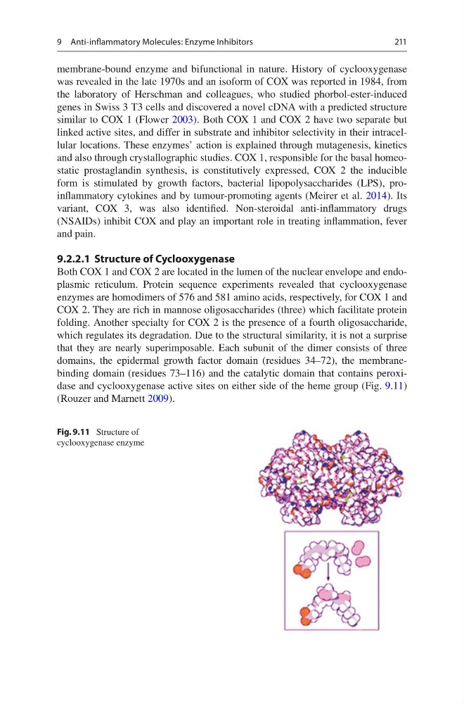
domains, the epidermal growth factor domain (residues 34–72), the membranebinding domain (residues 73–116) and the catalytic domain that contains peroxidase and cyclooxygenase active sites on either side of the heme group (Fig. 9.11)
(Rouzer and Marnett 2009).
Fig. 9.11 Structure of
cyclooxygenase enzyme
9 Anti-inflammatory Molecules: Enzyme Inhibitors
membrane- bound enzyme and bifunctional in nature. History of cyclooxygenase
was revealed in the late 1970s and an isoform of COX was reported in 1984, from
the laboratory of Herschman and colleagues, who studied phorbol-ester-induced
genes in Swiss 3 T3 cells and discovered a novel cDNA with a predicted structure
similar to COX 1 (Flower 2003). Both COX 1 and COX 2 have two separate but
linked active sites, and differ in substrate and inhibitor selectivity in their intracellular locations. These enzymes’ action is explained through mutagenesis, kinetics
and also through crystallographic studies. COX 1, responsible for the basal homeostatic prostaglandin synthesis, is constitutively expressed, COX 2 the inducible
form is stimulated by growth factors, bacterial lipopolysaccharides (LPS), proinflammatory cytokines and by tumour-promoting agents (Meirer et al. 2014). Its
variant, COX 3, was also identified. Non-steroidal anti-inflammatory drugs
(NSAIDs) inhibit COX and play an important role in treating inflammation, fever
and pain.
9.2.2.1 Structure of Cyclooxygenase
Both COX 1 and COX 2 are located in the lumen of the nuclear envelope and endoplasmic reticulum. Protein sequence experiments revealed that cyclooxygenase
enzymes are homodimers of 576 and 581 amino acids, respectively, for COX 1 and
COX 2. They are rich in mannose oligosaccharides (three) which facilitate protein
folding. Another specialty for COX 2 is the presence of a fourth oligosaccharide,
which regulates its degradation. Due to the structural similarity, it is not a surprise
that they are nearly superimposable. Each subunit of the dimer consists of three
domains, the epidermal growth factor domain (residues 34–72), the membranebinding domain (residues 73–116) and the catalytic domain that contains peroxidase and cyclooxygenase active sites on either side of the heme group (Fig. 9.11)
(Rouzer and Marnett 2009).
Fig. 9.11 Structure of
cyclooxygenase enzyme
9 Anti-inflammatory Molecules: Enzyme Inhibitors