

236
Mohamed A1-Rubeai
clear that light scatters cannot always provide adequate discrimination of
apoptosis and necrosis in heterogeneous cell cultures.
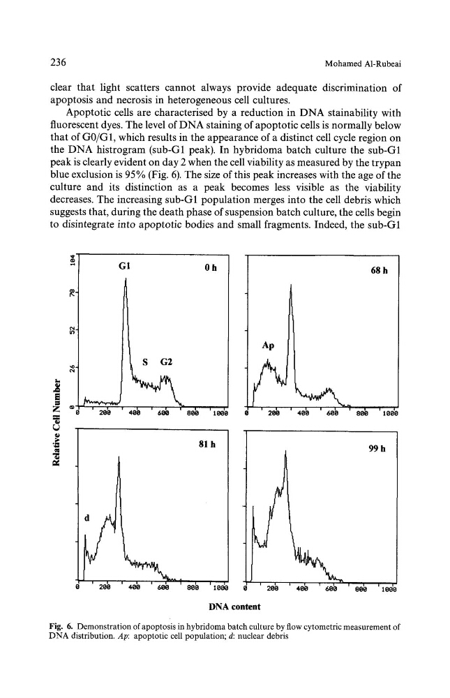
Apoptotic cells are characterised by a reduction in DNA stainability with
fluorescent dyes. The level of DNA staining of apoptotic cells is normally below
that of G0/G1, which results in the appearance of a distinct cell cycle region on
the DNA histrogram (sub-G1 peak). In hybridoma batch culture the sub-G1
peak is clearly evident on day 2 when the cell viability as measured by the trypan
blue exclusion is 95% (Fig. 6). The size of this peak increases with the age of the
culture and its distinction as a peak becomes less visible as the viability
decreases. The increasing sub-G1 population merges into the cell debris which
suggests that, during the death phase of suspension batch culture, the cells begin
to disintegrate into apoptotic bodies and small fragments. Indeed, the sub-G1
t...
z
qk
G1
0 h
i
i
i
|
i
i
0
20b
400
6o0
seb to00
81 h
68 h
' 200
400
600
000
d
' 200 ' 400 ' 600 ' 800 '1000
J
' 1000
' 20~
DNA content
400
600
99 h
e
e
00~ '10~o
Fig. 6. Demonstration of apoptosis in hybridoma batch culture by flow cytometric measurement of
DNA distribution. Ap: apoptotic cell population; d: nuclear debris
Mohamed A1-Rubeai
clear that light scatters cannot always provide adequate discrimination of
apoptosis and necrosis in heterogeneous cell cultures.
Apoptotic cells are characterised by a reduction in DNA stainability with
fluorescent dyes. The level of DNA staining of apoptotic cells is normally below
that of G0/G1, which results in the appearance of a distinct cell cycle region on
the DNA histrogram (sub-G1 peak). In hybridoma batch culture the sub-G1
peak is clearly evident on day 2 when the cell viability as measured by the trypan
blue exclusion is 95% (Fig. 6). The size of this peak increases with the age of the
culture and its distinction as a peak becomes less visible as the viability
decreases. The increasing sub-G1 population merges into the cell debris which
suggests that, during the death phase of suspension batch culture, the cells begin
to disintegrate into apoptotic bodies and small fragments. Indeed, the sub-G1
t...
z
qk
G1
0 h
i
i
i
|
i
i
0
20b
400
6o0
seb to00
81 h
68 h
' 200
400
600
000
d
' 200 ' 400 ' 600 ' 800 '1000
J
' 1000
' 20~
DNA content
400
600
99 h
e
e
00~ '10~o
Fig. 6. Demonstration of apoptosis in hybridoma batch culture by flow cytometric measurement of
DNA distribution. Ap: apoptotic cell population; d: nuclear debris