

Apoptosis and Cell Culture Technology
235
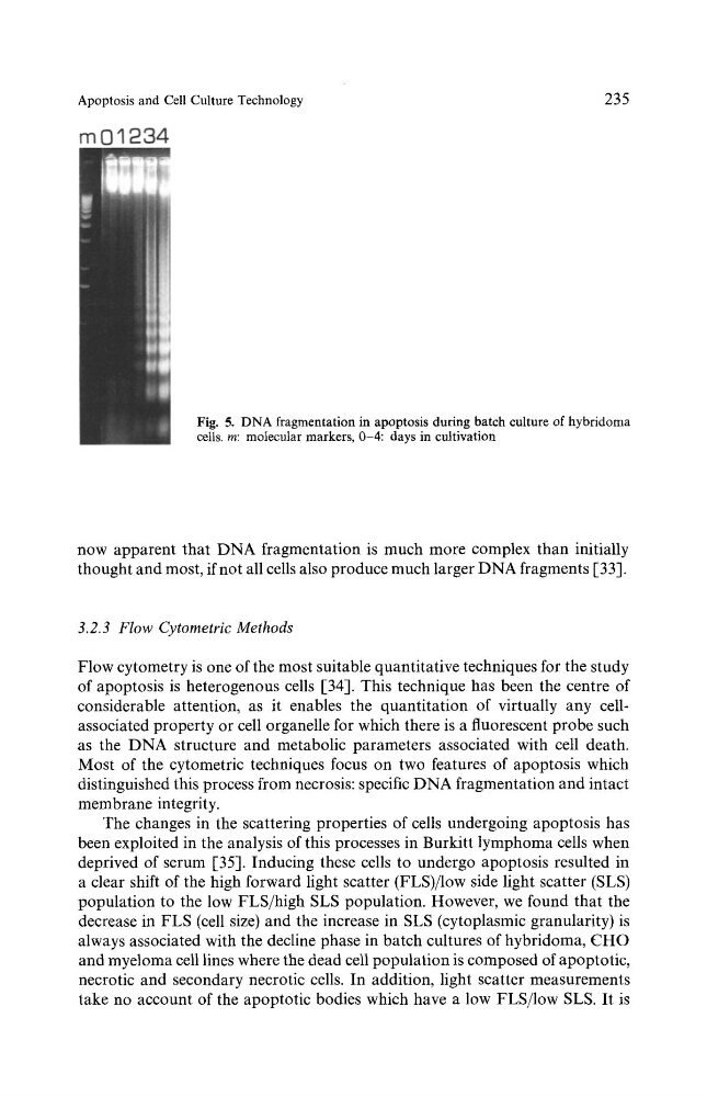
Fig. 5. DNA fragmentation in apoptosis during batch culture of hybridoma
cells, m: molecular markers, 0-4: days in cultivation
now apparent that DNA fragmentation is much more complex than initially
thought and most, if not all cells also produce much larger DNA fragments [33].
3.2.3 Flow Cytometric Methods
Flow cytometry is one of the most suitable quantitative techniques for the study
of apoptosis is heterogenous cells [-34]. This technique has been the centre of
considerable attention, as it enables the quantitation of virtually any cellassociated property or cell organelle for which there is a fluorescent probe such
as the DNA structure and metabolic parameters associated with cell death.
Most of the cytometric techniques focus on two features of apoptosis which
distinguished this process from necrosis: specific DNA fragmentation and intact
membrane integrity.
The changes in the scattering properties of cells undergoing apoptosis has
been exploited in the analysis of this processes in Burkitt lymphoma cells when
deprived of serum [-35]. Inducing these cells to undergo apoptosis resulted in
a clear shift of the high forward light scatter (FLS)/low side light scatter (SLS)
population to the low FLS/high SLS population. However, we found that the
decrease in FLS (cell size) and the increase in SLS (cytoplasmic granularity) is
always associated with the decline phase in batch cultures of hybridoma, CHO
and myeloma cell lines where the dead cell population is composed of apoptotic,
necrotic and secondary necrotic cells. In addition, light scatter measurements
take no account of the apoptotic bodies which have a low FLS/low SLS. It is
235
Fig. 5. DNA fragmentation in apoptosis during batch culture of hybridoma
cells, m: molecular markers, 0-4: days in cultivation
now apparent that DNA fragmentation is much more complex than initially
thought and most, if not all cells also produce much larger DNA fragments [33].
3.2.3 Flow Cytometric Methods
Flow cytometry is one of the most suitable quantitative techniques for the study
of apoptosis is heterogenous cells [-34]. This technique has been the centre of
considerable attention, as it enables the quantitation of virtually any cellassociated property or cell organelle for which there is a fluorescent probe such
as the DNA structure and metabolic parameters associated with cell death.
Most of the cytometric techniques focus on two features of apoptosis which
distinguished this process from necrosis: specific DNA fragmentation and intact
membrane integrity.
The changes in the scattering properties of cells undergoing apoptosis has
been exploited in the analysis of this processes in Burkitt lymphoma cells when
deprived of serum [-35]. Inducing these cells to undergo apoptosis resulted in
a clear shift of the high forward light scatter (FLS)/low side light scatter (SLS)
population to the low FLS/high SLS population. However, we found that the
decrease in FLS (cell size) and the increase in SLS (cytoplasmic granularity) is
always associated with the decline phase in batch cultures of hybridoma, CHO
and myeloma cell lines where the dead cell population is composed of apoptotic,
necrotic and secondary necrotic cells. In addition, light scatter measurements
take no account of the apoptotic bodies which have a low FLS/low SLS. It is