

the effective field strength due to the variation in its conductivity with temperature. In order to overcome this problem the strength of the phosphate
buffer is reduced from 0.01 mol l –1 to 0.002 mol l –1 . Note that the electrophoretic mobility of any given species increases with decreasing buffer ionic strength
[74]. This enhancement at lower concentration enables the separation of
cells/particles whose mobilities are only slightly different from each other. The
electrophoretic extraction results of latex particles with lower ionic strength
buffer are encouraging (data not show). As already discussed, the most useful
application of ADSEP electrophoresis is to fractionate mixed cells/particles,
exploiting the differences in their mobilities. The fractionation of a mixture
containing fixed blood cells and latex particles is to be carried out.
Calculations predicted heating rates of the order of millidegrees per
second for the applied current using a field that would affect motion of particles with electrophoretic mobilities around 10 –4 m 2 V –1 s –1 [72]. Theoretical
values of temperature-increase (predicted values) were calculated using
buffers of known conductivity and compared with exact measurements. In
0.01 mol l
–1 phosphate buffer, k E = 3.6 S m
–1 (which is considered a high
conductivity buffer for use in an electrophoretic instrument) a field of approximately 0.05 V m
–1 was applied [72]. The Joule-heating calculation with t
in seconds is given by
DT = (IE/ÇC p )t = 0.017 (t)
(17)
The plot depicting the above relationship, indicating the variation of DT with
the time of application of field at various values of electric currents, is shown in
Fig. 12. The above relationship was counter checked by measuring the temperature with a thermistor probe over a 2-min period (twice the typical time
period of one electrophoretic transfer) and obtaining the following linear
relationship:
DT = (1.2 °C/120) = 0.01(t)
(18)
Multistage Magnetic and Electrophoretic Extraction of Cells, Particles and Macromolecules
163
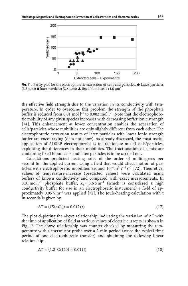
Fig. 11. Parity plot for the electrophoretic extraction of cells and particles. v Latex particles
(3.5 mm); latex particles (2.6 mm); ̆ fixed blood cells (4.6 mm)
buffer is reduced from 0.01 mol l –1 to 0.002 mol l –1 . Note that the electrophoretic mobility of any given species increases with decreasing buffer ionic strength
[74]. This enhancement at lower concentration enables the separation of
cells/particles whose mobilities are only slightly different from each other. The
electrophoretic extraction results of latex particles with lower ionic strength
buffer are encouraging (data not show). As already discussed, the most useful
application of ADSEP electrophoresis is to fractionate mixed cells/particles,
exploiting the differences in their mobilities. The fractionation of a mixture
containing fixed blood cells and latex particles is to be carried out.
Calculations predicted heating rates of the order of millidegrees per
second for the applied current using a field that would affect motion of particles with electrophoretic mobilities around 10 –4 m 2 V –1 s –1 [72]. Theoretical
values of temperature-increase (predicted values) were calculated using
buffers of known conductivity and compared with exact measurements. In
0.01 mol l
–1 phosphate buffer, k E = 3.6 S m
–1 (which is considered a high
conductivity buffer for use in an electrophoretic instrument) a field of approximately 0.05 V m
–1 was applied [72]. The Joule-heating calculation with t
in seconds is given by
DT = (IE/ÇC p )t = 0.017 (t)
(17)
The plot depicting the above relationship, indicating the variation of DT with
the time of application of field at various values of electric currents, is shown in
Fig. 12. The above relationship was counter checked by measuring the temperature with a thermistor probe over a 2-min period (twice the typical time
period of one electrophoretic transfer) and obtaining the following linear
relationship:
DT = (1.2 °C/120) = 0.01(t)
(18)
Multistage Magnetic and Electrophoretic Extraction of Cells, Particles and Macromolecules
163
Fig. 11. Parity plot for the electrophoretic extraction of cells and particles. v Latex particles
(3.5 mm); latex particles (2.6 mm); ̆ fixed blood cells (4.6 mm)