

48
HPLC
Flow
IOO-1000ullmln
Prototype DAD flow cell
onhne LC-ESI-MSIMS
Teflon sleeve
IllS' 0 0 250 m 10
1. CASTELLANOS-SERRA et al.
One piece nut
Fused SIlica
,../
.. '
1/16- 0 D
178um I D
Spilt-Tee .:
....
;.......
. ...
Fused Silica tubing
PEEK tulllng
Sample
collector
f
MIcro flow
SOum Peek tubing
Mlcrocolumn
Excess flow
Sample Inlecto,
Sample loop
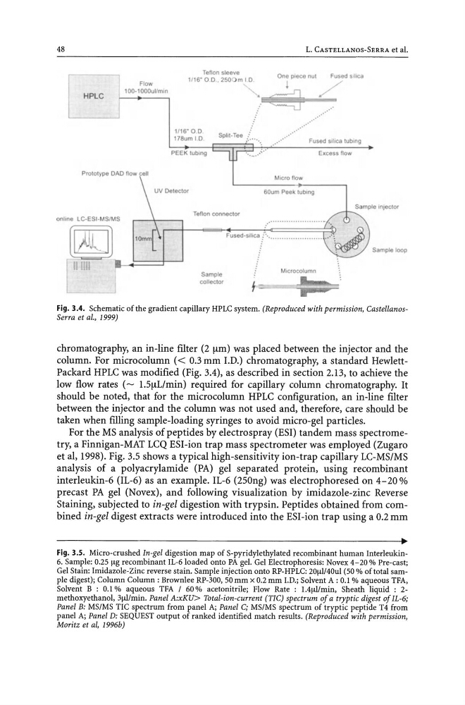
Fig. 3.4. Schematic of the gradient capillary HPLC system. (Reproduced with permission, CastellanosSerra et al., 1999)
chromatography, an in-line fIlter (2 !-tm) was placed between the injector and the
column. For microcolumn « 0.3 mm I.D.) chromatography, a standard HewlettPackard HPLC was modified (Fig. 3.4), as described in section 2.13, to achieve the
low flow rates (~ 1.5!-tLlmin) required for capillary column chromatography. It
should be noted, that for the micro column HPLC configuration, an in-line filter
between the injector and the column was not used and, therefore, care should be
taken when filling sample-loading syringes to avoid micro-gel particles.
For the MS analysis of peptides by electro spray (ESI) tandem mass spectrometry, a Finnigan-MAT LCQ ESI-ion trap mass spectrometer was employed (Zugaro
et aI, 1998). Fig. 3.5 shows a typical high-sensitivity ion-trap capillary LC-MS/MS
analysis of a polyacrylamide (PA) gel separated protein, using recombinant
interleukin-6 (IL-6) as an example. IL-6 (250ng) was electrophoresed on 4-20 %
precast PA gel (Novex), and following visualization by imidazole-zinc Reverse
Staining, subjected to in-gel digestion with trypsin. Pep tides obtained from combined in-gel digest extracts were introduced into the ESI-ion trap using a 0.2 mm
Fig. 3.S. Micro-crushed In-gel digestion map of S-pyridylethylated recombinant human Interleukin6. Sample: 0.25 Ilg recombinant IL-6loaded onto PA gel. Gel Electrophoresis: Novex 4- 20% Pre-cast;
Gel Stain: Imidazole-Zinc reverse stain. Sample injection onto RP-HPLC: 20ll1/40ul (50 % of total sample digest); Column Column: Brownlee RP-300, 50 mm x 0.2 mm l.D.; Solvent A: 0.1 % aqueous TFA,
Solvent B : 0.1 % aqueous TFA I 60 % acetonitrile; Flow Rate : 1.41l1/min, Sheath liquid : 2methoxyethanol, 31ll/min. Panel A:xKU> Total-ion-current (TIC) spectrum of a tryptic digest of IL-6;
Panel B: MS/MS TIC spectrum from panel A; Panel C; MS/MS spectrum of tryptic peptide T4 from
panel A; Panel D: SEQUEST output of ranked identified match results. (Reproduced with permission,
Moritz et ai, 1996b)
HPLC
Flow
IOO-1000ullmln
Prototype DAD flow cell
onhne LC-ESI-MSIMS
Teflon sleeve
IllS' 0 0 250 m 10
1. CASTELLANOS-SERRA et al.
One piece nut
Fused SIlica
,../
.. '
1/16- 0 D
178um I D
Spilt-Tee .:
....
;.......
. ...
Fused Silica tubing
PEEK tulllng
Sample
collector
f
MIcro flow
SOum Peek tubing
Mlcrocolumn
Excess flow
Sample Inlecto,
Sample loop
Fig. 3.4. Schematic of the gradient capillary HPLC system. (Reproduced with permission, CastellanosSerra et al., 1999)
chromatography, an in-line fIlter (2 !-tm) was placed between the injector and the
column. For microcolumn « 0.3 mm I.D.) chromatography, a standard HewlettPackard HPLC was modified (Fig. 3.4), as described in section 2.13, to achieve the
low flow rates (~ 1.5!-tLlmin) required for capillary column chromatography. It
should be noted, that for the micro column HPLC configuration, an in-line filter
between the injector and the column was not used and, therefore, care should be
taken when filling sample-loading syringes to avoid micro-gel particles.
For the MS analysis of peptides by electro spray (ESI) tandem mass spectrometry, a Finnigan-MAT LCQ ESI-ion trap mass spectrometer was employed (Zugaro
et aI, 1998). Fig. 3.5 shows a typical high-sensitivity ion-trap capillary LC-MS/MS
analysis of a polyacrylamide (PA) gel separated protein, using recombinant
interleukin-6 (IL-6) as an example. IL-6 (250ng) was electrophoresed on 4-20 %
precast PA gel (Novex), and following visualization by imidazole-zinc Reverse
Staining, subjected to in-gel digestion with trypsin. Pep tides obtained from combined in-gel digest extracts were introduced into the ESI-ion trap using a 0.2 mm
Fig. 3.S. Micro-crushed In-gel digestion map of S-pyridylethylated recombinant human Interleukin6. Sample: 0.25 Ilg recombinant IL-6loaded onto PA gel. Gel Electrophoresis: Novex 4- 20% Pre-cast;
Gel Stain: Imidazole-Zinc reverse stain. Sample injection onto RP-HPLC: 20ll1/40ul (50 % of total sample digest); Column Column: Brownlee RP-300, 50 mm x 0.2 mm l.D.; Solvent A: 0.1 % aqueous TFA,
Solvent B : 0.1 % aqueous TFA I 60 % acetonitrile; Flow Rate : 1.41l1/min, Sheath liquid : 2methoxyethanol, 31ll/min. Panel A:xKU> Total-ion-current (TIC) spectrum of a tryptic digest of IL-6;
Panel B: MS/MS TIC spectrum from panel A; Panel C; MS/MS spectrum of tryptic peptide T4 from
panel A; Panel D: SEQUEST output of ranked identified match results. (Reproduced with permission,
Moritz et ai, 1996b)