

332
• .. I t O ~'
l - fr
•
i
. .
"'"
. .,,,,,
"''''11\''''
U"''''' I»
pott . 61 l , .. belolt
... JIII r"oPIM"
r - " - - - --'
i~
"II'
i
" ...
...
ii'
P. M. RUDD et al.
" ,
. I"' llKo\ldo u
. [I lI,l(Q~lIdo 'il
~I t Sl,oDl,d!l 1f
• ~JoI I v(Oill)) ~
. (t hlu~C:-!l l t
· &1 Q l1.octo l llhnr
;.:.,1 U"" III:~ U
_ ~1001 I II,o'.o!G tt
• Cl tIlCO$'i!O u
• 81 ~'2lClltlo , ·.:t:J 1I
• Sf ~ f ' OI
IIlau
it
I . . ... """ ..
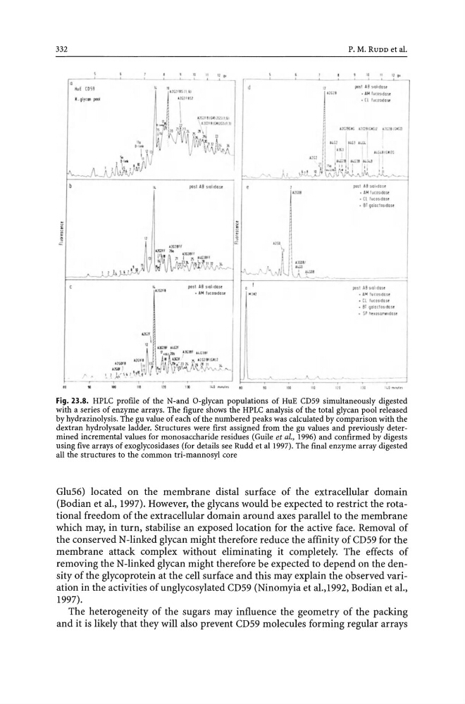
Fig. 23.8. HPLC profile of the N-and O-glycan populations of HuE CD59 simultaneously digested
with a series of enzyme arrays. The figure shows the HPLC analysis of the total glycan pool released
by hydrazinolysis. The gu value of each of the numbered peaks was calculated by comparison with the
dextran hydrolysate ladder. Structures were first assigned from the gu values and previously determined incremental values for monosaccharide residues (Guile et a/., 1996) and confirmed by digests
using five arrays of exoglycosidases (for details see Rudd et aI1997). The final enzyme array digested
all the structures to the common tri-mannosyl core
Glu56) located on the membrane distal surface of the extracellular domain
(Bodian et al., 1997). However, the glycans would be expected to restrict the rotational freedom of the extracellular domain around axes parallel to the membrane
which may, in turn, stabilise an exposed location for the active face. Removal of
the conserved N-linked glycan might therefore reduce the affinity of CD 59 for the
membrane attack complex without eliminating it completely. The effects of
removing the N-linked glycan might therefore be expected to depend on the density of the glycoprotein at the cell surface and this may explain the observed variation in the activities of unglycosylated CD59 (Ninomyia et aI.,1992, Bodian et aI.,
1997).
The heterogeneity of the sugars may influence the geometry of the packing
and it is likely that they will also prevent CD59 molecules forming regular arrays
• .. I t O ~'
l - fr
•
i
. .
"'"
. .,,,,,
"''''11\''''
U"''''' I»
pott . 61 l , .. belolt
... JIII r"oPIM"
r - " - - - --'
i~
"II'
i
" ...
...
ii'
P. M. RUDD et al.
" ,
. I"' llKo\ldo u
. [I lI,l(Q~lIdo 'il
~I t Sl,oDl,d!l 1f
• ~JoI I v(Oill)) ~
. (t hlu~C:-!l l t
· &1 Q l1.octo l llhnr
;.:.,1 U"" III:~ U
_ ~1001 I II,o'.o!G tt
• Cl tIlCO$'i!O u
• 81 ~'2lClltlo , ·.:t:J 1I
• Sf ~ f ' OI
IIlau
it
I . . ... """ ..
Fig. 23.8. HPLC profile of the N-and O-glycan populations of HuE CD59 simultaneously digested
with a series of enzyme arrays. The figure shows the HPLC analysis of the total glycan pool released
by hydrazinolysis. The gu value of each of the numbered peaks was calculated by comparison with the
dextran hydrolysate ladder. Structures were first assigned from the gu values and previously determined incremental values for monosaccharide residues (Guile et a/., 1996) and confirmed by digests
using five arrays of exoglycosidases (for details see Rudd et aI1997). The final enzyme array digested
all the structures to the common tri-mannosyl core
Glu56) located on the membrane distal surface of the extracellular domain
(Bodian et al., 1997). However, the glycans would be expected to restrict the rotational freedom of the extracellular domain around axes parallel to the membrane
which may, in turn, stabilise an exposed location for the active face. Removal of
the conserved N-linked glycan might therefore reduce the affinity of CD 59 for the
membrane attack complex without eliminating it completely. The effects of
removing the N-linked glycan might therefore be expected to depend on the density of the glycoprotein at the cell surface and this may explain the observed variation in the activities of unglycosylated CD59 (Ninomyia et aI.,1992, Bodian et aI.,
1997).
The heterogeneity of the sugars may influence the geometry of the packing
and it is likely that they will also prevent CD59 molecules forming regular arrays