

328
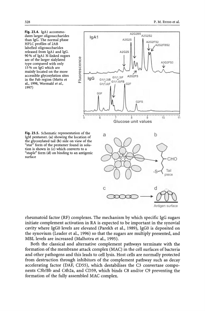
Fig. 23.4. IgAI accommodates larger oligo saccharides
than IgG. The normal phase
HPLC profiles of 2AB
labelled oligosaccharides
released from IgAI and IgG.
90 % of IgAI N-linked sugars
are of the larger sialylated
type compared with only
Q)
u
c
Q)
u
(/)
IgA1
P. M. RUDD et al.
A2G2:Z1G2BSA2G2S2
A2G2FS 2
~ , A2G2 FBS2
A2G2S
1
15 % on IgG which are
mainly located on the more
accessible glycosylation sites
in the Fab region (Mattu et
al., 1998, Wormald et aI.,
1997)
~
~.J\--J'--_ ___ -
g i G
Gl (1, 3)F A2G2FS
u..
9
Gl (P )B Gl(1:6)FB
Gl( 1,6)F J G2F
GOF
1·········1·" ····'·1'··,· · "·1·······'·1········ · 1··· . . . ... ,
5
6
7
B
9
1 0
11
Fig. 23.S. Schematic representation of the
IgM pentamer. (a) showing the location of
the glycosylated tail (b) side on view of the
"star" form of the pentamer found in solution is shown in (c) which converts to a
"staple" form (d) on binding to an antigenic
surface
Glucose unit values
a
b
c d .. ·.
oo:x:o - . d(J\J
Antigen surface
rheumatoid factor (RF) complexes. The mechanism by which specific IgG sugars
initiate complement activation in RA is expected to be important in the synovial
cavity where IgGO levels are elevated (Parekh et al., 1989), IgGO is deposited on
the synovium (Leader et al., 1996) so that the sugars are multiply presented, and
MBL levels are increased (Malhotra et al., 1995).
Both the classical and alternative complement pathways terminate with the
formation of the membrane attack complex (MAC) in the cell surfaces of bacteria
and other pathogens and this leads to cell lysis. Host cells are normally protected
from destruction through inhibitors of the complement pathway such as decay
accelerating factor (DAF, CD55), which destabilises the C3 convertase components C3b/Bb and C4b2a, and CD59, which binds C8 and/or C9 preventing the
formation of the fully assembled MAC complex.
Fig. 23.4. IgAI accommodates larger oligo saccharides
than IgG. The normal phase
HPLC profiles of 2AB
labelled oligosaccharides
released from IgAI and IgG.
90 % of IgAI N-linked sugars
are of the larger sialylated
type compared with only
Q)
u
c
Q)
u
(/)
IgA1
P. M. RUDD et al.
A2G2:Z1G2BSA2G2S2
A2G2FS 2
~ , A2G2 FBS2
A2G2S
1
15 % on IgG which are
mainly located on the more
accessible glycosylation sites
in the Fab region (Mattu et
al., 1998, Wormald et aI.,
1997)
~
~.J\--J'--_ ___ -
g i G
Gl (1, 3)F A2G2FS
u..
9
Gl (P )B Gl(1:6)FB
Gl( 1,6)F J G2F
GOF
1·········1·" ····'·1'··,· · "·1·······'·1········ · 1··· . . . ... ,
5
6
7
B
9
1 0
11
Fig. 23.S. Schematic representation of the
IgM pentamer. (a) showing the location of
the glycosylated tail (b) side on view of the
"star" form of the pentamer found in solution is shown in (c) which converts to a
"staple" form (d) on binding to an antigenic
surface
Glucose unit values
a
b
c d .. ·.
oo:x:o - . d(J\J
Antigen surface
rheumatoid factor (RF) complexes. The mechanism by which specific IgG sugars
initiate complement activation in RA is expected to be important in the synovial
cavity where IgGO levels are elevated (Parekh et al., 1989), IgGO is deposited on
the synovium (Leader et al., 1996) so that the sugars are multiply presented, and
MBL levels are increased (Malhotra et al., 1995).
Both the classical and alternative complement pathways terminate with the
formation of the membrane attack complex (MAC) in the cell surfaces of bacteria
and other pathogens and this leads to cell lysis. Host cells are normally protected
from destruction through inhibitors of the complement pathway such as decay
accelerating factor (DAF, CD55), which destabilises the C3 convertase components C3b/Bb and C4b2a, and CD59, which binds C8 and/or C9 preventing the
formation of the fully assembled MAC complex.