

Exploring Functions for Glycosylation in Host Defence
323
which allows direct quantitation from HPLC profiles. In the HPLC technology a
column containing amide-silica resolves both charged and neutral N- and 0glycans in a single run. The glycans are separated on the basis of differences in
hydrophilicity, which reflects arm specificity, linkage and monosaccharide composition in a single run. The system uses a volatile buffer system so that individual glycans can be recovered free from buffer salts for analysis by mass spectrometry. The elution positions of the glycans, determined in glucose units with refer1
•
,
6 .
3
1 2
1 2 1
2 6 1 l
• 2
6 1 "
2 6 , "
2 a 1
1 "
• Mannose
• N-acetyl glucosam lne
*: Fucose
!:;, Galactose
<> Sia li c aCid
Q)
<.>
" Q)
<.>
III
~
0
"
iL
Sequencing IgG glycans
5
6 7 8 9 10 12 gu
I
I
I
I
I
I
I I I
IgG gly"n
G I F IG2F
POOl
I *-6 :J;~
GOF
/:rG2FS
\ G2fS
iJ }G2FBS
AB Sial
j02f
~
' 1
AS 51,,1
9T 1:1;)1
~F~
AB Sill I
9T ill
S~ hell
r
M3N2F
An ~I.l1
aT g3 1
SP he~
Clluc
?
80
90
100
110
120 130
minu te s
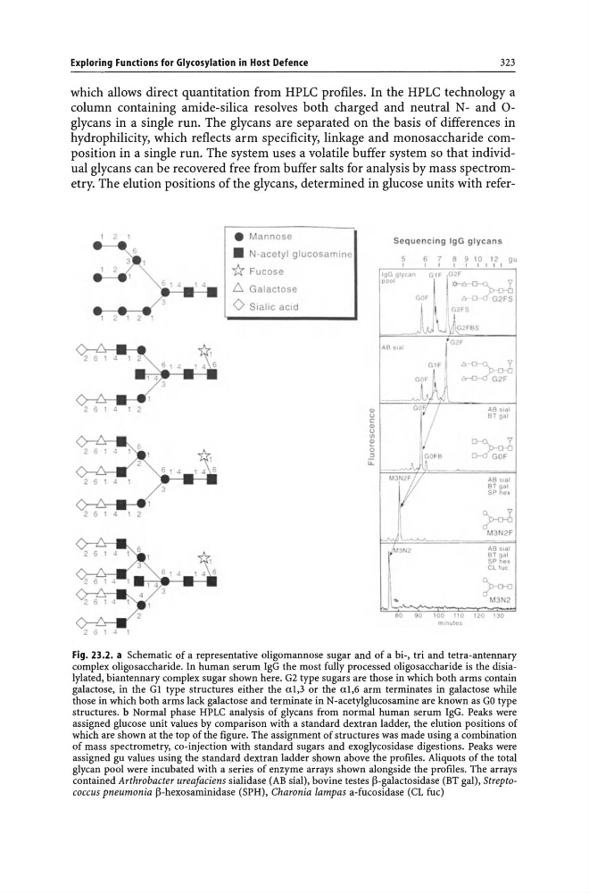
Fig. 23.2. a Schematic of a representative oligomannose sugar and of a bi-, tri and tetra-antennary
complex oligosaccharide. In human serum IgG the most fully processed oligosaccharide is the disialylated, biantennary complex sugar shown here. G2 type sugars are those in which both arms contain
galactose, in the Gl type structures either the al,3 or the al,6 arm terminates in galactose while
those in which both arms lack galactose and terminate in N -acetylglucosamine are known as GO type
structures. b Normal phase HPLC analysis of glycans from normal human serum IgG. Peaks were
assigned glucose unit values by comparison with a standard dextran ladder, the elution positions of
which are shown at the top of the figure. The assignment of structures was made using a combination
of mass spectrometry, co-injection with standard sugars and exoglycosidase digestions. Peaks were
assigned gu values using the standard dextran ladder shown above the profiles. Aliquots of the total
glycan pool were incubated with a series of enzyme arrays shown alongside the profiles. The arrays
contained Arthrobacter ureafaciens sialidase (AB sial), bovine testes j3-galactosidase (BT gal), Streptococcus pneumonia j3-hexosaminidase (SPH), Charonia lampas a-fucosidase (CL fuc)
323
which allows direct quantitation from HPLC profiles. In the HPLC technology a
column containing amide-silica resolves both charged and neutral N- and 0glycans in a single run. The glycans are separated on the basis of differences in
hydrophilicity, which reflects arm specificity, linkage and monosaccharide composition in a single run. The system uses a volatile buffer system so that individual glycans can be recovered free from buffer salts for analysis by mass spectrometry. The elution positions of the glycans, determined in glucose units with refer1
•
,
6 .
3
1 2
1 2 1
2 6 1 l
• 2
6 1 "
2 6 , "
2 a 1
1 "
• Mannose
• N-acetyl glucosam lne
*: Fucose
!:;, Galactose
<> Sia li c aCid
Q)
<.>
" Q)
<.>
III
~
0
"
iL
Sequencing IgG glycans
5
6 7 8 9 10 12 gu
I
I
I
I
I
I
I I I
IgG gly"n
G I F IG2F
POOl
I *-6 :J;~
GOF
/:rG2FS
\ G2fS
iJ }G2FBS
AB Sial
j02f
~
' 1
AS 51,,1
9T 1:1;)1
~F~
AB Sill I
9T ill
S~ hell
r
M3N2F
An ~I.l1
aT g3 1
SP he~
Clluc
?
80
90
100
110
120 130
minu te s
Fig. 23.2. a Schematic of a representative oligomannose sugar and of a bi-, tri and tetra-antennary
complex oligosaccharide. In human serum IgG the most fully processed oligosaccharide is the disialylated, biantennary complex sugar shown here. G2 type sugars are those in which both arms contain
galactose, in the Gl type structures either the al,3 or the al,6 arm terminates in galactose while
those in which both arms lack galactose and terminate in N -acetylglucosamine are known as GO type
structures. b Normal phase HPLC analysis of glycans from normal human serum IgG. Peaks were
assigned glucose unit values by comparison with a standard dextran ladder, the elution positions of
which are shown at the top of the figure. The assignment of structures was made using a combination
of mass spectrometry, co-injection with standard sugars and exoglycosidase digestions. Peaks were
assigned gu values using the standard dextran ladder shown above the profiles. Aliquots of the total
glycan pool were incubated with a series of enzyme arrays shown alongside the profiles. The arrays
contained Arthrobacter ureafaciens sialidase (AB sial), bovine testes j3-galactosidase (BT gal), Streptococcus pneumonia j3-hexosaminidase (SPH), Charonia lampas a-fucosidase (CL fuc)