

266
R. E. HARRINGTON et al.
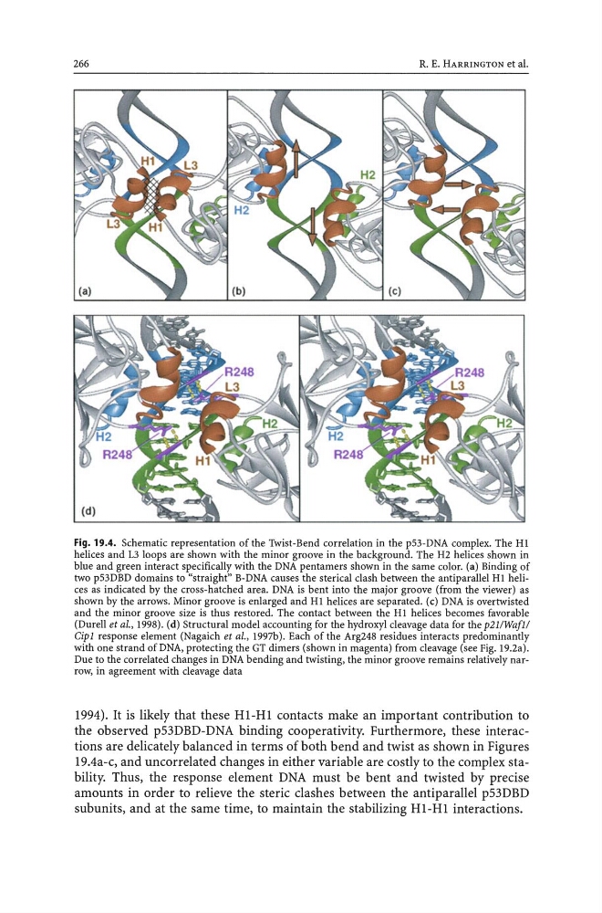
Fig. 19.4. Schematic representation of the Twist-Bend correlation in the p53-DNA complex. The HI
helices and L3 loops are shown with the minor groove in the background. The H2 helices shown in
blue and green interact specifically with the DNA pentamers shown in the same color. (a) Binding of
two p53DBD domains to "straight" B-DNA causes the sterical clash between the antiparallel HI helices as indicated by the cross-hatched area. DNA is bent into the major groove (from the viewer) as
shown by the arrows. Minor groove is enlarged and HI helices are separated. (c) DNA is overtwisted
and the minor groove size is thus restored. The contact between the HI helices becomes favorable
(Durell et aI., 1998). (d) Structural model accounting for the hydroxyl cleavage data for the p2I!WafI!
Cipl response element (Nagaich et aI., 1997b). Each of the Arg248 residues interacts predominantly
with one strand of DNA, protecting the GT dimers (shown in magenta) from cleavage (see Fig. 19.2a).
Due to the correlated changes in DNA bending and twisting, the minor groove remains relatively narrow, in agreement with cleavage data
1994). It is likely that these HI-HI contacts make an important contribution to
the observed p53DBD-DNA binding cooperativity. Furthermore, these interactions are delicately balanced in terms of both bend and twist as shown in Figures
19.4a-c, and uncorrelated changes in either variable are costly to the complex stability. Thus, the response element DNA must be bent and twisted by precise
amounts in order to relieve the steric clashes between the antiparallel p53DBD
subunits, and at the same time, to maintain the stabilizing HI-HI interactions.
R. E. HARRINGTON et al.
Fig. 19.4. Schematic representation of the Twist-Bend correlation in the p53-DNA complex. The HI
helices and L3 loops are shown with the minor groove in the background. The H2 helices shown in
blue and green interact specifically with the DNA pentamers shown in the same color. (a) Binding of
two p53DBD domains to "straight" B-DNA causes the sterical clash between the antiparallel HI helices as indicated by the cross-hatched area. DNA is bent into the major groove (from the viewer) as
shown by the arrows. Minor groove is enlarged and HI helices are separated. (c) DNA is overtwisted
and the minor groove size is thus restored. The contact between the HI helices becomes favorable
(Durell et aI., 1998). (d) Structural model accounting for the hydroxyl cleavage data for the p2I!WafI!
Cipl response element (Nagaich et aI., 1997b). Each of the Arg248 residues interacts predominantly
with one strand of DNA, protecting the GT dimers (shown in magenta) from cleavage (see Fig. 19.2a).
Due to the correlated changes in DNA bending and twisting, the minor groove remains relatively narrow, in agreement with cleavage data
1994). It is likely that these HI-HI contacts make an important contribution to
the observed p53DBD-DNA binding cooperativity. Furthermore, these interactions are delicately balanced in terms of both bend and twist as shown in Figures
19.4a-c, and uncorrelated changes in either variable are costly to the complex stability. Thus, the response element DNA must be bent and twisted by precise
amounts in order to relieve the steric clashes between the antiparallel p53DBD
subunits, and at the same time, to maintain the stabilizing HI-HI interactions.