

A Structural Model for the PS3 Complex with DNA Response Elements
265
than one allosterically related conformational state of the entire protein molecule
(Vojtesek et aI., 1995; Waterman et aI., 1995). It may also rationalize the apparent
discrepancy between the observations that
(i) wild type p53 can bind to "split" response elements containing various
amounts of spacer DNA (Cook et al., 1995), which in some cases may be
functional (Tokino et al., 1994), and that
(ii) p53DBD binds to split sites only if the spacers contain an integral number of
helical turns of DNA and if both half sites incorporate the flexible tetrad
CATG at the pentameric junctions (P. Balagurumoorthy et aI., manuscript
submitted).
The reason that the tetrameric model requires a coupling of bending and twisting
is illustrated in Fig. 19.4. When the p53DBD subunits are bound to the "straight"
B-DNA, the antiparallel HI-helices produce unacceptable steric clashes (Fig.
19.4a). One of the ways to relieve these clashes is to bend the DNA into the major
groove (Fig. 19.4b). In such a case, however, the HI-HI interactions can be lost
completely, which is also unfavorable. Energy calculations (Durell et aI., 1998)
indicate that the optimum is achieved when the DNA is both bent and twisted,
and the two HI helices are closely juxtaposed, forming a two helix bundle as
shown in Fig. 19.4c. Each antiparallel dimer in the tetrameric complex is stabilized by van der Waals interactions and hydrogen bonds involving His 178, Glu180
and Arg181 (Durell et aI., 1998). Notably, the asymmetric interactions of Arg248
with the DNA backbone predicted by this model (Fig. 19.4d), are consistent with
the OH radical protection results (Nagaich et aI., 1997b). This is not apparent
from the co-crystal structure in which only a single p53DBD is bound to the DNA
and the Arg248 residue is symmetrically bound in the minor groove (Cho et aI.,
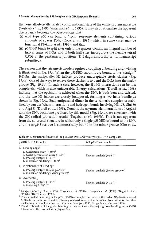
Table 19.1. Structural features of the p53DBD-DNA and wild type p53-DNA complexes
p53DBD-DNA Complex
WT p53-DNA complex
A. Bending angle$
1. Cyclization assay (~60 0) 1
2. Cyclic permutation assay (~50 0)2
3. Phasing analysis (~35 0)3
4. Molecular modeling (~40 0)4
Phasing analysis (~55 0)3
B. Directionality of Bending'
1. Phasing analysis (Major groove)3
2. Molecular modeling (Major groove)4
Phasing analysis (Major groove) 3
C. Overtwisting
1. Phasing analysis (~35 0) 3
2. Modeling (~25 0)3,5
Phasing analysis (~70 0) 3
1 Balagurumoorthy et al. (1995). 2Nagaich et al. (1997a). 3Nagaich et al. (1999). 4 Nagaich et al.
(1997b). 5Durell et al. (1998).
$ The estimated bend angles for p53DBD-DNA complex decrease in the order: (Cyclization assay)
> (Cyclic permutation assay) > (Phasing analysis), in accord with earlier observation for the other
nucleoprotein complexes (Van der Vliet and Verrijzer, 1993; Kerppola and Curran, 1993).
* The directionality of the global bending is consistent with the major groove bending in the CATG
tetramers in the two half sites (Figure 2c).
265
than one allosterically related conformational state of the entire protein molecule
(Vojtesek et aI., 1995; Waterman et aI., 1995). It may also rationalize the apparent
discrepancy between the observations that
(i) wild type p53 can bind to "split" response elements containing various
amounts of spacer DNA (Cook et al., 1995), which in some cases may be
functional (Tokino et al., 1994), and that
(ii) p53DBD binds to split sites only if the spacers contain an integral number of
helical turns of DNA and if both half sites incorporate the flexible tetrad
CATG at the pentameric junctions (P. Balagurumoorthy et aI., manuscript
submitted).
The reason that the tetrameric model requires a coupling of bending and twisting
is illustrated in Fig. 19.4. When the p53DBD subunits are bound to the "straight"
B-DNA, the antiparallel HI-helices produce unacceptable steric clashes (Fig.
19.4a). One of the ways to relieve these clashes is to bend the DNA into the major
groove (Fig. 19.4b). In such a case, however, the HI-HI interactions can be lost
completely, which is also unfavorable. Energy calculations (Durell et aI., 1998)
indicate that the optimum is achieved when the DNA is both bent and twisted,
and the two HI helices are closely juxtaposed, forming a two helix bundle as
shown in Fig. 19.4c. Each antiparallel dimer in the tetrameric complex is stabilized by van der Waals interactions and hydrogen bonds involving His 178, Glu180
and Arg181 (Durell et aI., 1998). Notably, the asymmetric interactions of Arg248
with the DNA backbone predicted by this model (Fig. 19.4d), are consistent with
the OH radical protection results (Nagaich et aI., 1997b). This is not apparent
from the co-crystal structure in which only a single p53DBD is bound to the DNA
and the Arg248 residue is symmetrically bound in the minor groove (Cho et aI.,
Table 19.1. Structural features of the p53DBD-DNA and wild type p53-DNA complexes
p53DBD-DNA Complex
WT p53-DNA complex
A. Bending angle$
1. Cyclization assay (~60 0) 1
2. Cyclic permutation assay (~50 0)2
3. Phasing analysis (~35 0)3
4. Molecular modeling (~40 0)4
Phasing analysis (~55 0)3
B. Directionality of Bending'
1. Phasing analysis (Major groove)3
2. Molecular modeling (Major groove)4
Phasing analysis (Major groove) 3
C. Overtwisting
1. Phasing analysis (~35 0) 3
2. Modeling (~25 0)3,5
Phasing analysis (~70 0) 3
1 Balagurumoorthy et al. (1995). 2Nagaich et al. (1997a). 3Nagaich et al. (1999). 4 Nagaich et al.
(1997b). 5Durell et al. (1998).
$ The estimated bend angles for p53DBD-DNA complex decrease in the order: (Cyclization assay)
> (Cyclic permutation assay) > (Phasing analysis), in accord with earlier observation for the other
nucleoprotein complexes (Van der Vliet and Verrijzer, 1993; Kerppola and Curran, 1993).
* The directionality of the global bending is consistent with the major groove bending in the CATG
tetramers in the two half sites (Figure 2c).