

A Structural Model for the PS3 Complex with DNA Response Elements
259
2
Structure of the p53-DNA Nucleoprotein Complex
2.1
Tetrameric Nature of the p53 Complex
Wild type p53 binds as a tetramer (El-Deiry et al., 1992; Friedman et al., 1993;
Halazonetis and Kandil, 1993; Wang et al., 1994) to over 100 different naturally
occurring response elements of which approximately 60 exhibit functionality. It
has been estimated that the human genome contains approximately 200 to 300
such sites (Tokino et al., 1994). Response elements differ in details of specific
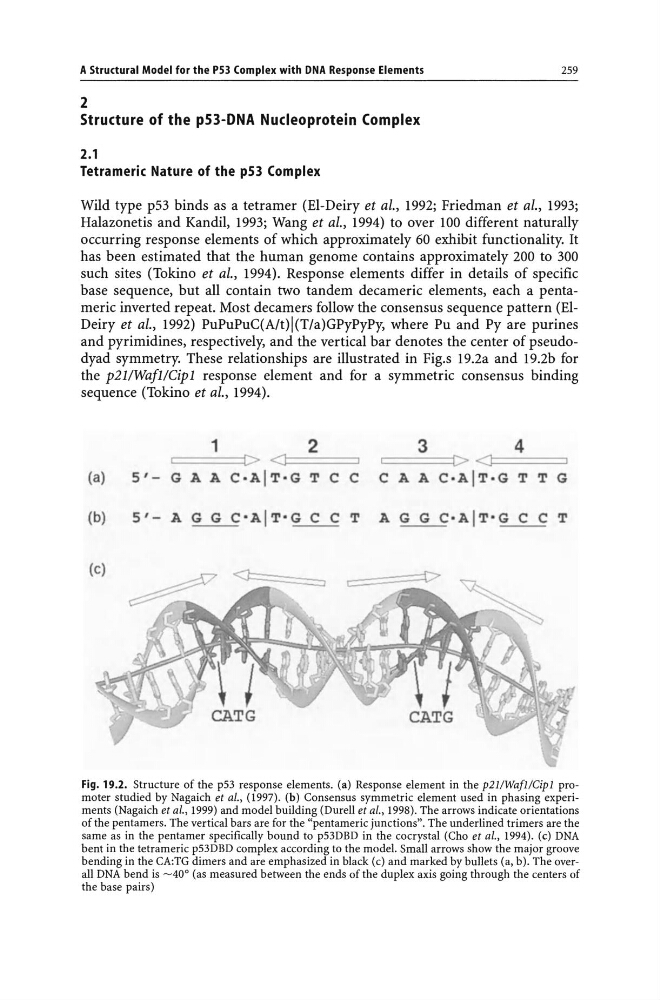
base sequence, but all contain two tandem decameric elements, each a pentameric inverted repeat. Most decamers follow the consensus sequence pattern (ElDeiry et al., 1992) PuPuPuC(A/t)I(T/a)GPyPyPy, where Pu and Py are purines
and pyrimidines, respectively, and the vertical bar denotes the center of pseudodyad symmetry. These relationships are illustrated in Fig.s 19.2a and 19.2b for
the p211WafllCipl response element and for a symmetric consensus binding
sequence (Tokino et al., 1994).
(a)
(b)
1
2
====:::j[> <11=:==
5'- G A A C.AIT.G Tee
5'- A G G c'AIT'G C C T
3
4
===[><1===
C A A C.AIT.G T T G
A G G c.AIT·G C C T
Fig. 19.2. Structure of the p53 response elements. (a) Response element in the p2I1WafIlCipl promoter studied by Nagaich et aI., (1997). (b) Consensus symmetric element used in phasing experiments (Nagaich et al., 1999) and model building (Durell et aI., 1998). The arrows indicate orientations
of the pentamers. The vertical bars are for the "pentameric junctions". The underlined trimers are the
same as in the pentamer specifically bound to p53DBD in the cocrystal (Cho et aI., 1994). (e) DNA
bent in the tetrameric p53DBD complex according to the model. Small arrows show the major groove
bending in the CA:TG dimers and are emphasized in black (c) and marked by bullets (a, b). The overall DNA bend is -40 0 (as measured between the ends of the duplex axis going through the centers of
the base pairs)
259
2
Structure of the p53-DNA Nucleoprotein Complex
2.1
Tetrameric Nature of the p53 Complex
Wild type p53 binds as a tetramer (El-Deiry et al., 1992; Friedman et al., 1993;
Halazonetis and Kandil, 1993; Wang et al., 1994) to over 100 different naturally
occurring response elements of which approximately 60 exhibit functionality. It
has been estimated that the human genome contains approximately 200 to 300
such sites (Tokino et al., 1994). Response elements differ in details of specific
base sequence, but all contain two tandem decameric elements, each a pentameric inverted repeat. Most decamers follow the consensus sequence pattern (ElDeiry et al., 1992) PuPuPuC(A/t)I(T/a)GPyPyPy, where Pu and Py are purines
and pyrimidines, respectively, and the vertical bar denotes the center of pseudodyad symmetry. These relationships are illustrated in Fig.s 19.2a and 19.2b for
the p211WafllCipl response element and for a symmetric consensus binding
sequence (Tokino et al., 1994).
(a)
(b)
1
2
====:::j[> <11=:==
5'- G A A C.AIT.G Tee
5'- A G G c'AIT'G C C T
3
4
===[><1===
C A A C.AIT.G T T G
A G G c.AIT·G C C T
Fig. 19.2. Structure of the p53 response elements. (a) Response element in the p2I1WafIlCipl promoter studied by Nagaich et aI., (1997). (b) Consensus symmetric element used in phasing experiments (Nagaich et al., 1999) and model building (Durell et aI., 1998). The arrows indicate orientations
of the pentamers. The vertical bars are for the "pentameric junctions". The underlined trimers are the
same as in the pentamer specifically bound to p53DBD in the cocrystal (Cho et aI., 1994). (e) DNA
bent in the tetrameric p53DBD complex according to the model. Small arrows show the major groove
bending in the CA:TG dimers and are emphasized in black (c) and marked by bullets (a, b). The overall DNA bend is -40 0 (as measured between the ends of the duplex axis going through the centers of
the base pairs)