

258
R. E. HARRINGTON et al.
Wild type p53 contains at least four functional domains. A schematic of the
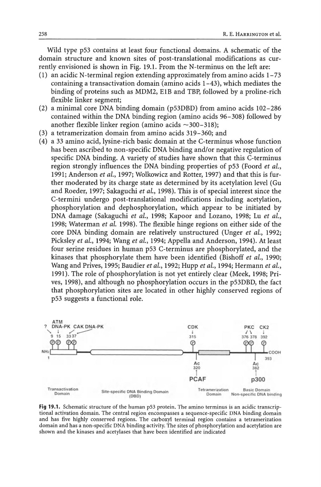
domain structure and known sites of post-translational modifications as currently envisioned is shown in Fig. 19.1. From the N-terminus on the left are:
(1) an acidic N-terminal region extending approximately from amino acids 1-73
containing a transactivation domain (amino acids 1-43), which mediates the
binding of proteins such as MDM2, EIB and TBP, followed by a proline-rich
flexible linker segment;
(2) a minimal core DNA binding domain (p53DBD) from amino acids 102-286
contained within the DNA binding region (amino acids 96-308) followed by
another flexible linker region (amino acids -300-318);
(3) a tetramerization domain from amino acids 319-360; and
(4) a 33 amino acid, lysine-rich basic domain at the C-terminus whose function
has been ascribed to non-specific DNA binding and/or negative regulation of
specific DNA binding. A variety of studies have shown that this C-terminus
region strongly influences the DNA binding properties of p53 (Foord et al.,
1991; Anderson et al., 1997; Wolkowicz and Rotter, 1997) and that this is further moderated by its charge state as determined by its acetylation level (Gu
and Roeder, 1997; Sakaguchi et ai., 1998). This is of special interest since the
C-termini undergo post-translational modifications including acetylation,
phosphorylation and dephosphorylation, which appear to be initiated by
DNA damage (Sakaguchi et ai., 1998; Kapoor and Lozano, 1998; Lu et ai.,
1998; Waterman et al. 1998). The flexible hinge regions on either side of the
core DNA binding domain are relatively unstructured (Unger et al., 1992;
Picksley et ai., 1994; Wang et al., 1994; Appella and Anderson, 1994). At least
four serine residues in human p53 C-terminus are phosphorylated, and the
kinases that phosphorylate them have been identified (Bishoff et al., 1990;
Wang and Prives, 1995; Baudier et al., 1992; Hupp et al., 1994; Hermann et al.,
1991). The role of phosphorylation is not yet entirely clear (Meek, 1998; Prives, 1998), and although no phosphorylation occurs in the p53DBD, the fact
that phosphorylation sites are located in other highly conserved regions of
p53 suggests a functional role.
ATM
? DNA·PK CAK DNA· PK
CDK
PKC CK2
"
j
/...---I
1\
j
9 15 3337
315
376378 392
?T ~
J1 1.
n f
N H 2C
~~ ______________________ ~.
.~ ~:1t= . J~I2 ~rJ--~----Lrl--l- COOH
393
Transactiva tion
Domai n
Si t e ~ speel f ic DN A Bindi ng Domain
(DBD)
Ac
320
1
PCAF
Ac
382
1
p300
Tctramcrizalion
Basic Oom ai n
Doma in
Non-specific DN A bi nding
Fig 19.1. Schematic structure of the human p53 protein. The amino terminus is an acidic transcriptional activation domain. The central region encompasses a sequence-specific DNA binding domain
and has five highly conserved regions. The carboxyl terminal region contains a tetramerization
domain and has a non-specific DNA binding activity. The sites of phosphorylation and acetylation are
shown and the kinases and acetylases that have been identified are indicated
R. E. HARRINGTON et al.
Wild type p53 contains at least four functional domains. A schematic of the
domain structure and known sites of post-translational modifications as currently envisioned is shown in Fig. 19.1. From the N-terminus on the left are:
(1) an acidic N-terminal region extending approximately from amino acids 1-73
containing a transactivation domain (amino acids 1-43), which mediates the
binding of proteins such as MDM2, EIB and TBP, followed by a proline-rich
flexible linker segment;
(2) a minimal core DNA binding domain (p53DBD) from amino acids 102-286
contained within the DNA binding region (amino acids 96-308) followed by
another flexible linker region (amino acids -300-318);
(3) a tetramerization domain from amino acids 319-360; and
(4) a 33 amino acid, lysine-rich basic domain at the C-terminus whose function
has been ascribed to non-specific DNA binding and/or negative regulation of
specific DNA binding. A variety of studies have shown that this C-terminus
region strongly influences the DNA binding properties of p53 (Foord et al.,
1991; Anderson et al., 1997; Wolkowicz and Rotter, 1997) and that this is further moderated by its charge state as determined by its acetylation level (Gu
and Roeder, 1997; Sakaguchi et ai., 1998). This is of special interest since the
C-termini undergo post-translational modifications including acetylation,
phosphorylation and dephosphorylation, which appear to be initiated by
DNA damage (Sakaguchi et ai., 1998; Kapoor and Lozano, 1998; Lu et ai.,
1998; Waterman et al. 1998). The flexible hinge regions on either side of the
core DNA binding domain are relatively unstructured (Unger et al., 1992;
Picksley et ai., 1994; Wang et al., 1994; Appella and Anderson, 1994). At least
four serine residues in human p53 C-terminus are phosphorylated, and the
kinases that phosphorylate them have been identified (Bishoff et al., 1990;
Wang and Prives, 1995; Baudier et al., 1992; Hupp et al., 1994; Hermann et al.,
1991). The role of phosphorylation is not yet entirely clear (Meek, 1998; Prives, 1998), and although no phosphorylation occurs in the p53DBD, the fact
that phosphorylation sites are located in other highly conserved regions of
p53 suggests a functional role.
ATM
? DNA·PK CAK DNA· PK
CDK
PKC CK2
"
j
/...---I
1\
j
9 15 3337
315
376378 392
?T ~
J1 1.
n f
N H 2C
~~ ______________________ ~.
.~ ~:1t= . J~I2 ~rJ--~----Lrl--l- COOH
393
Transactiva tion
Domai n
Si t e ~ speel f ic DN A Bindi ng Domain
(DBD)
Ac
320
1
PCAF
Ac
382
1
p300
Tctramcrizalion
Basic Oom ai n
Doma in
Non-specific DN A bi nding
Fig 19.1. Schematic structure of the human p53 protein. The amino terminus is an acidic transcriptional activation domain. The central region encompasses a sequence-specific DNA binding domain
and has five highly conserved regions. The carboxyl terminal region contains a tetramerization
domain and has a non-specific DNA binding activity. The sites of phosphorylation and acetylation are
shown and the kinases and acetylases that have been identified are indicated