

Structural Characterisation of Porcine Seminal Plasma Psp-I/Psp-1i
247
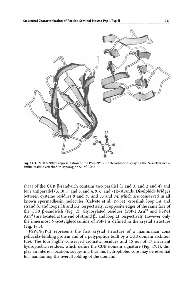
Fig. 17.3. MOLSCRIPT representation of the PSP-I/PSP-I! heterodimer displaying the N-acetylglucosamine residue attached to asparagine 50 of PSP-I
sheet of the CUB ~-sandwich contains two parallel (1 and 3, and 2 and 4) and
four antiparallel (3, 10, 5, and 8, and 4, 9, 6, and 7) ~-strands. Disulphide bridges
between cysteine residues 9 and 30 and 53 and 74, which are conserved in all
known spermadhesin molecules (Calvete et al. 1995a), crosslink loop LA and
strand ~4 and loops LE and LG, respectively, at opposite edges of the same face of
the CUB ~-sandwich (Fig. 2). Glycosylated residues (PSP-I Asn 50 and PSP-II
Asn 98 ) are located at the end of strand ~5 and loop 11, respectively. However, only
the innermost N -acetylglucosamine of PSP-I is defined in the crystal structure
(Fig. 17.3).
PSP-I/PSP-II represents the first crystal structure of a mammalian zona
pellucida-binding protein and of a polypeptide built by a CUB domain architecture. The four highly conserved aromatic residues and 15 out of 17 invariant
hydrophobic residues, which define the CUB domain signature (Fig. 17.1), display an interior location, suggesting that this hydrophobic core may be essential
for maintaining the overall folding of the domain.
247
Fig. 17.3. MOLSCRIPT representation of the PSP-I/PSP-I! heterodimer displaying the N-acetylglucosamine residue attached to asparagine 50 of PSP-I
sheet of the CUB ~-sandwich contains two parallel (1 and 3, and 2 and 4) and
four antiparallel (3, 10, 5, and 8, and 4, 9, 6, and 7) ~-strands. Disulphide bridges
between cysteine residues 9 and 30 and 53 and 74, which are conserved in all
known spermadhesin molecules (Calvete et al. 1995a), crosslink loop LA and
strand ~4 and loops LE and LG, respectively, at opposite edges of the same face of
the CUB ~-sandwich (Fig. 2). Glycosylated residues (PSP-I Asn 50 and PSP-II
Asn 98 ) are located at the end of strand ~5 and loop 11, respectively. However, only
the innermost N -acetylglucosamine of PSP-I is defined in the crystal structure
(Fig. 17.3).
PSP-I/PSP-II represents the first crystal structure of a mammalian zona
pellucida-binding protein and of a polypeptide built by a CUB domain architecture. The four highly conserved aromatic residues and 15 out of 17 invariant
hydrophobic residues, which define the CUB domain signature (Fig. 17.1), display an interior location, suggesting that this hydrophobic core may be essential
for maintaining the overall folding of the domain.