

246
J. J. CALVETE and L. SANZ
sperm-egg recognition, the large amino acid sequence identity shared with any
other spermadhesin molecule (Fig. 17.1) indicates that the conformation ofPSP-I
(and PSP-II) may be highly conserved in other spermadhesins. In addition, the
carbohydrate- and zona pellucida glycoprotein-binding capabilities of PSP-II
makes this spermadhesin a paradigm molecule for the whole protein family.
Interestingly, besides its potential importance in mammalian fertilisation,
spermadhesins are interesting targets for structure elucidation because this
group of proteins is built by a single CUB domain architecture (Bork, Beckmann
1993). The CUB domain is a novell00-llO-residue module first reported in complement subcomponents ~lr/Cls, embryonic sea urchin protein lIegf, and bone
morphogenetic protein 1 mmpl). CUB domains have been subsequently identified in 16 functionally diverse proteins, many of which are known to be involved
in developmental processes (Bork, Beckmann 1993). Analysis of conserved structural features in a multiple sequence alignment of CUB domains revealed the
presence of several rather conserved blocks interrupted by variable regions of
flexible length. Conserved characteristics include the four cysteine residues,
which in spermadhesins have been shown to form disulphide bridges between
neighbour cysteine residues, and various conserved hydrophobic and aromatic
positions.
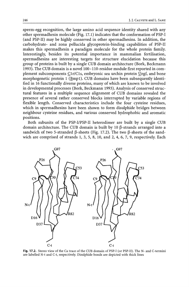
Both subunits of the PSP-I/PSP-II heterodimer are built by a single CUB
domain architecture. The CUB domain is built by 10 ~-strands arranged into a
sandwich of two 5-stranded ~-sheets (Fig. 17.2). The two ~-sheets of the sandwich are comprised of strands 1, 3, 5, 8, 10, and 2, 4, 6, 7, 9, respectively. Each
G97
N-t
N-t
C-t
C-t
Fig. 17.2. Stereo view of the Ca trace of the CUB domain ofPSP-I (or PSP-II). The N- and C-termini
are labelled N-t and C-t, respectively. Disulphide bonds are depicted with thick lines
J. J. CALVETE and L. SANZ
sperm-egg recognition, the large amino acid sequence identity shared with any
other spermadhesin molecule (Fig. 17.1) indicates that the conformation ofPSP-I
(and PSP-II) may be highly conserved in other spermadhesins. In addition, the
carbohydrate- and zona pellucida glycoprotein-binding capabilities of PSP-II
makes this spermadhesin a paradigm molecule for the whole protein family.
Interestingly, besides its potential importance in mammalian fertilisation,
spermadhesins are interesting targets for structure elucidation because this
group of proteins is built by a single CUB domain architecture (Bork, Beckmann
1993). The CUB domain is a novell00-llO-residue module first reported in complement subcomponents ~lr/Cls, embryonic sea urchin protein lIegf, and bone
morphogenetic protein 1 mmpl). CUB domains have been subsequently identified in 16 functionally diverse proteins, many of which are known to be involved
in developmental processes (Bork, Beckmann 1993). Analysis of conserved structural features in a multiple sequence alignment of CUB domains revealed the
presence of several rather conserved blocks interrupted by variable regions of
flexible length. Conserved characteristics include the four cysteine residues,
which in spermadhesins have been shown to form disulphide bridges between
neighbour cysteine residues, and various conserved hydrophobic and aromatic
positions.
Both subunits of the PSP-I/PSP-II heterodimer are built by a single CUB
domain architecture. The CUB domain is built by 10 ~-strands arranged into a
sandwich of two 5-stranded ~-sheets (Fig. 17.2). The two ~-sheets of the sandwich are comprised of strands 1, 3, 5, 8, 10, and 2, 4, 6, 7, 9, respectively. Each
G97
N-t
N-t
C-t
C-t
Fig. 17.2. Stereo view of the Ca trace of the CUB domain ofPSP-I (or PSP-II). The N- and C-termini
are labelled N-t and C-t, respectively. Disulphide bonds are depicted with thick lines