

228
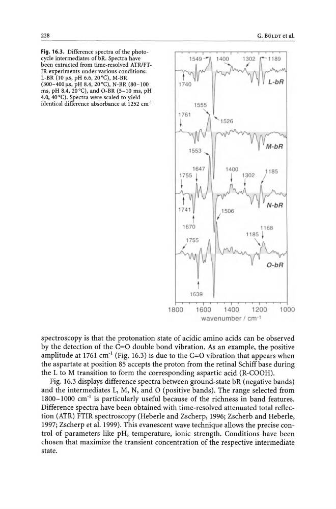
Fig. 16.3. Difference spectra of the photocycle intermediates of bR. Spectra have
been extracted from time-resolved ATRlFTIR experiments under various conditions:
L-BR (lOlls, pH 6.6, 20°C), M-BR
(300-400 1lS, pH 8.4, 20°C), N-BR (80-100
ms, pH 8.4, 20°C), and O-BR (5-10 ms, pH
4.0, 40°C). Spectra were scaled to yield
identical difference absorbance at 1252 cm· l
G. BULDT et al.
1185
t
/
1506
1670
1639
1800
1600
1400
1200
1000
wavenumber I em' !
spectroscopy is that the protonation state of acidic amino acids can be observed
by the detection of the c=o double bond vibration. As an example, the positive
amplitude at 1761 cm· l (Fig. 16.3) is due to the c=o vibration that appears when
the aspartate at position 85 accepts the proton from the retinal Schiff base during
the L to M transition to form the corresponding aspartic acid (R-COOH).
Fig. 16.3 displays difference spectra between ground-state bR (negative bands)
and the intermediates L, M, N, and 0 (positive bands). The range selected from
1800-1000 cm· l is particularly useful because of the richness in band features.
Difference spectra have been obtained with time-resolved attenuated total reflection (ATR) FTIR spectroscopy (Heberle and Zscherp, 1996; Zscherb and Heberle,
1997; Zscherp et al. 1999). This evanescent wave technique allows the precise control of parameters like pH, temperature, ionic strength. Conditions have been
chosen that maximize the transient concentration of the respective intermediate
state.
Fig. 16.3. Difference spectra of the photocycle intermediates of bR. Spectra have
been extracted from time-resolved ATRlFTIR experiments under various conditions:
L-BR (lOlls, pH 6.6, 20°C), M-BR
(300-400 1lS, pH 8.4, 20°C), N-BR (80-100
ms, pH 8.4, 20°C), and O-BR (5-10 ms, pH
4.0, 40°C). Spectra were scaled to yield
identical difference absorbance at 1252 cm· l
G. BULDT et al.
1185
t
/
1506
1670
1639
1800
1600
1400
1200
1000
wavenumber I em' !
spectroscopy is that the protonation state of acidic amino acids can be observed
by the detection of the c=o double bond vibration. As an example, the positive
amplitude at 1761 cm· l (Fig. 16.3) is due to the c=o vibration that appears when
the aspartate at position 85 accepts the proton from the retinal Schiff base during
the L to M transition to form the corresponding aspartic acid (R-COOH).
Fig. 16.3 displays difference spectra between ground-state bR (negative bands)
and the intermediates L, M, N, and 0 (positive bands). The range selected from
1800-1000 cm· l is particularly useful because of the richness in band features.
Difference spectra have been obtained with time-resolved attenuated total reflection (ATR) FTIR spectroscopy (Heberle and Zscherp, 1996; Zscherb and Heberle,
1997; Zscherp et al. 1999). This evanescent wave technique allows the precise control of parameters like pH, temperature, ionic strength. Conditions have been
chosen that maximize the transient concentration of the respective intermediate
state.