

226
EI61
0104
0102
Ret
'. .,
038
036
lTlOplasmic sic/"
D96
-'---- K216
~--- D85
extracell"lar ~ide
G. BiiLDT et al.
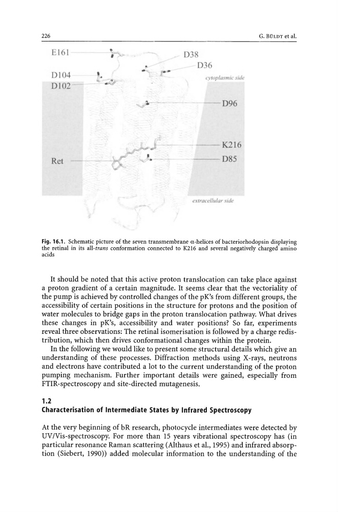
Fig. 16.1. Schematic picture of the seven transmembrane a-helices of bacteriorhodopsin displaying
the retinal in its all-trans conformation connected to K216 and several negatively charged amino
acids
It should be noted that this active proton translocation can take place against
a proton gradient of a certain magnitude. It seems clear that the vectoriality of
the pump is achieved by controlled changes of the pK's from different groups, the
accessibility of certain positions in the structure for protons and the position of
water molecules to bridge gaps in the proton translocation pathway. What drives
these changes in pK's, accessibility and water positions? So far, experiments
reveal three observations: The retinal isomerisation is followed by a charge redistribution, which then drives conformational changes within the protein.
In the following we would like to present some structural details which give an
understanding of these processes. Diffraction methods using X-rays, neutrons
and electrons have contributed a lot to the current understanding of the proton
pumping mechanism. Further important details were gained, especially from
FTIR-spectroscopy and site-directed mutagenesis.
1.2
Characterisation of Intermediate States by Infrared Spectroscopy
At the very beginning of bR research, photocycle intermediates were detected by
UVlVis-spectroscopy. For more than 15 years vibrational spectroscopy has (in
particular resonance Raman scattering (Althaus et aI., 1995) and infrared absorption (Siebert, 1990» added molecular information to the understanding of the
EI61
0104
0102
Ret
'. .,
038
036
lTlOplasmic sic/"
D96
-'---- K216
~--- D85
extracell"lar ~ide
G. BiiLDT et al.
Fig. 16.1. Schematic picture of the seven transmembrane a-helices of bacteriorhodopsin displaying
the retinal in its all-trans conformation connected to K216 and several negatively charged amino
acids
It should be noted that this active proton translocation can take place against
a proton gradient of a certain magnitude. It seems clear that the vectoriality of
the pump is achieved by controlled changes of the pK's from different groups, the
accessibility of certain positions in the structure for protons and the position of
water molecules to bridge gaps in the proton translocation pathway. What drives
these changes in pK's, accessibility and water positions? So far, experiments
reveal three observations: The retinal isomerisation is followed by a charge redistribution, which then drives conformational changes within the protein.
In the following we would like to present some structural details which give an
understanding of these processes. Diffraction methods using X-rays, neutrons
and electrons have contributed a lot to the current understanding of the proton
pumping mechanism. Further important details were gained, especially from
FTIR-spectroscopy and site-directed mutagenesis.
1.2
Characterisation of Intermediate States by Infrared Spectroscopy
At the very beginning of bR research, photocycle intermediates were detected by
UVlVis-spectroscopy. For more than 15 years vibrational spectroscopy has (in
particular resonance Raman scattering (Althaus et aI., 1995) and infrared absorption (Siebert, 1990» added molecular information to the understanding of the