

4 In Situ Nick End-Labeling: Electron Microscopical
41
/
granular keralinocyte
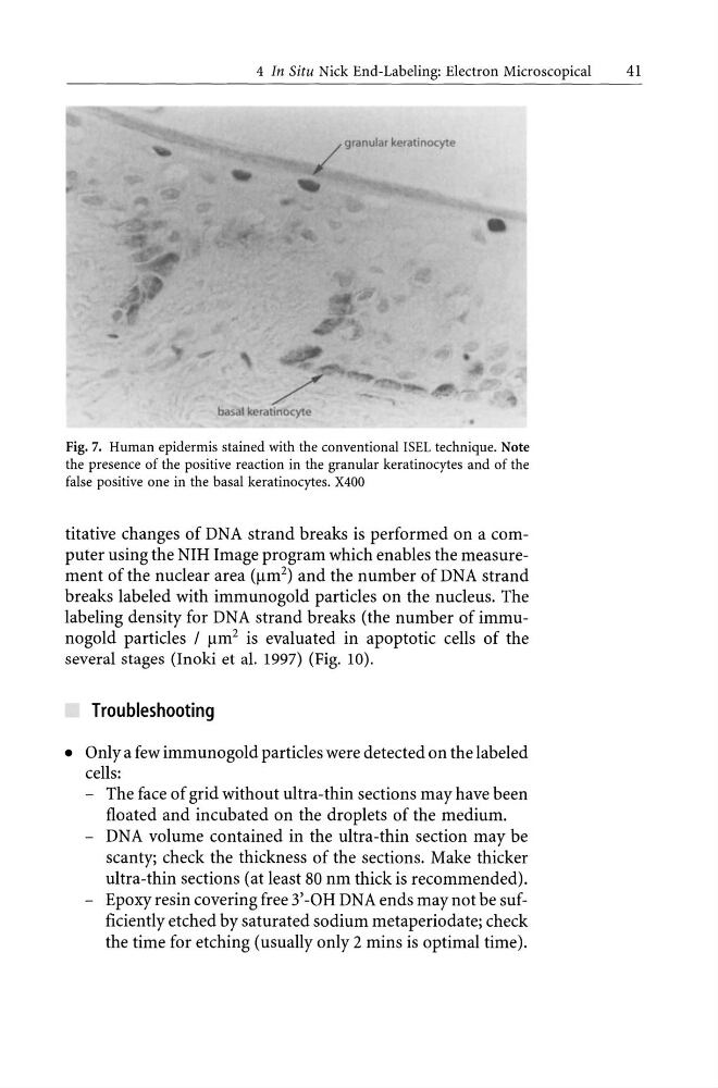
Fig. 7. Human epidermis stained with the conventional ISEL technique. Note
the presence of the positive reaction in the granular keratinocytes and of the
false positive one in the basal keratinocytes. X400
titative changes of DNA strand breaks is performed on a computer using the NIH Image program which enables the measurement of the nuclear area ()lm 2 ) and the number of DNA strand
breaks labeled with immunogold particles on the nucleus. The
labeling density for DNA strand breaks (the number of immuno gold particles / )lm 2 is evaluated in apoptotic cells of the
several stages (Inoki et al. 1997) (Fig. 10).
Troubleshooting
• Only a few immunogold particles were detected on the labeled
cells:
- The face of grid without ultra-thin sections may have been
floated and incubated on the droplets of the medium.
- DNA volume contained in the ultra-thin section may be
scanty; check the thickness of the sections. Make thicker
ultra-thin sections (at least 80 nm thick is recommended).
- Epoxy resin covering free 3' -OH DNA ends may not be sufficiently etched by saturated sodium metaperiodate; check
the time for etching (usually only 2 mins is optimal time).
41
/
granular keralinocyte
Fig. 7. Human epidermis stained with the conventional ISEL technique. Note
the presence of the positive reaction in the granular keratinocytes and of the
false positive one in the basal keratinocytes. X400
titative changes of DNA strand breaks is performed on a computer using the NIH Image program which enables the measurement of the nuclear area ()lm 2 ) and the number of DNA strand
breaks labeled with immunogold particles on the nucleus. The
labeling density for DNA strand breaks (the number of immuno gold particles / )lm 2 is evaluated in apoptotic cells of the
several stages (Inoki et al. 1997) (Fig. 10).
Troubleshooting
• Only a few immunogold particles were detected on the labeled
cells:
- The face of grid without ultra-thin sections may have been
floated and incubated on the droplets of the medium.
- DNA volume contained in the ultra-thin section may be
scanty; check the thickness of the sections. Make thicker
ultra-thin sections (at least 80 nm thick is recommended).
- Epoxy resin covering free 3' -OH DNA ends may not be sufficiently etched by saturated sodium metaperiodate; check
the time for etching (usually only 2 mins is optimal time).