

15 Electron Microscopic in Situ Hybridization and its Combination
215
Postembedding method
Hybridization signals for rat GH mRNA are localized on the
polysomes of the rough endoplasmic reticulum using 20 nm
streptavidin gold (Fig. 8). Electron microscopic in situ hybridization with a sense probe for rat GH mRNA shows no hybridization signals (Fig. 9).
Combined immunohistochemistry and preembedding
in situ hybridization at an electron microscopic level
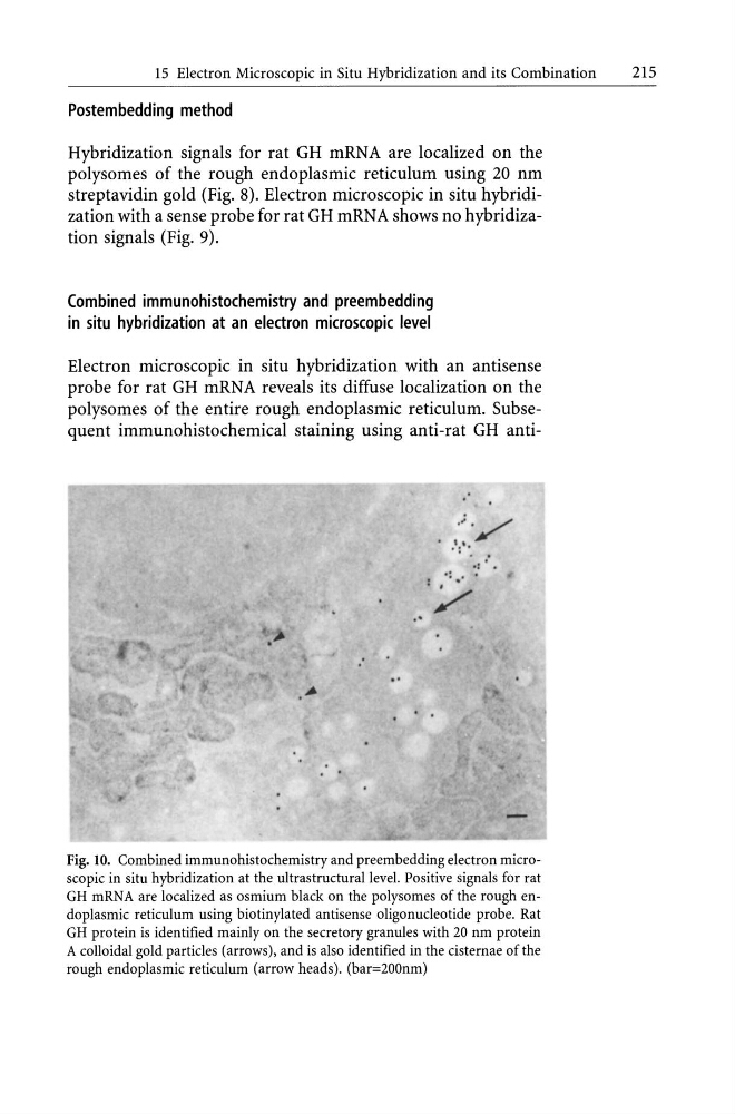
Electron microscopic in situ hybridization with an antisense
probe for rat GH mRNA reveals its diffuse localization on the
polysomes of the entire rough endoplasmic reticulum. Subsequent immunohistochemical staining using anti-rat GH anti. '
.'./
I ,
.: .
. :
.:." .
Fig. 10. Combined immunohistochemistry and preembedding electron microscopic in situ hybridization at the ultrastructural level. Positive signals for rat
GH mRNA are localized as osmium black on the polysomes of the rough endoplasmic reticulum using biotinylated antisense oligonucleotide probe. Rat
GH protein is identified mainly on the secretory granules with 20 nm protein
A colloidal gold particles (arrows), and is also identified in the cisternae of the
rough endoplasmic reticulum (arrow heads). (bar=200nm)
215
Postembedding method
Hybridization signals for rat GH mRNA are localized on the
polysomes of the rough endoplasmic reticulum using 20 nm
streptavidin gold (Fig. 8). Electron microscopic in situ hybridization with a sense probe for rat GH mRNA shows no hybridization signals (Fig. 9).
Combined immunohistochemistry and preembedding
in situ hybridization at an electron microscopic level
Electron microscopic in situ hybridization with an antisense
probe for rat GH mRNA reveals its diffuse localization on the
polysomes of the entire rough endoplasmic reticulum. Subsequent immunohistochemical staining using anti-rat GH anti. '
.'./
I ,
.: .
. :
.:." .
Fig. 10. Combined immunohistochemistry and preembedding electron microscopic in situ hybridization at the ultrastructural level. Positive signals for rat
GH mRNA are localized as osmium black on the polysomes of the rough endoplasmic reticulum using biotinylated antisense oligonucleotide probe. Rat
GH protein is identified mainly on the secretory granules with 20 nm protein
A colloidal gold particles (arrows), and is also identified in the cisternae of the
rough endoplasmic reticulum (arrow heads). (bar=200nm)