

180
YUKA IWASAKI AND IHACHI KATO
Results
To confirm the specificity of mRNA signals obtained by in situ
hybridization, a variety of control experiments are required. In
every run, sense and 28S rRNA probe are used as negative and
positive control, respectively. To exclude possible involvement
of proteins and DNA in signal formation, some sections are digested with RNase A (100 Ilg/ml) at 37°C for 1 hr, before hybridization. Others are hybridized with antisense probe in the presence of an excess amount of unlabeled antisense or sense oligoDNA, while some are washed under extremely high stringent
conditions after hybridization, to provide definitive evidence
for the sequence specificity of the signals.
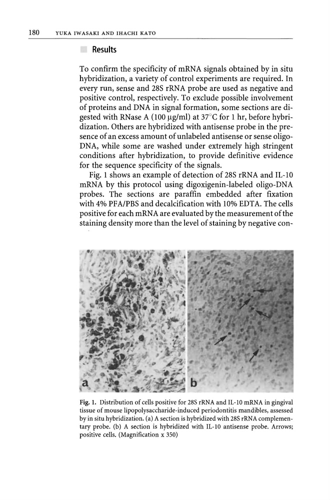
Fig. 1 shows an example of detection of 28S rRNA and IL-lO
mRNA by this protocol using digoxigenin-Iabeled oligo-DNA
probes. The sections are paraffin embedded after fixation
with 4% PF A/PBS and decalcification with 10% EDT A. The cells
positive for each mRN A are evaluated by the measurement of the
staining density more than the level of staining by negative conFig. 1. Distribution of cells positive for 28S rRNA and IL-10 mRNA in gingival
tissue of mouse lipopolysaccharide-induced periodontitis mandibles, assessed
by in situ hybridization. (a) A section is hybridized with 28S rRNA complementary probe. (b) A section is hybridized with IL-lO antisense probe. Arrows;
positive cells. (Magnification x 350)
YUKA IWASAKI AND IHACHI KATO
Results
To confirm the specificity of mRNA signals obtained by in situ
hybridization, a variety of control experiments are required. In
every run, sense and 28S rRNA probe are used as negative and
positive control, respectively. To exclude possible involvement
of proteins and DNA in signal formation, some sections are digested with RNase A (100 Ilg/ml) at 37°C for 1 hr, before hybridization. Others are hybridized with antisense probe in the presence of an excess amount of unlabeled antisense or sense oligoDNA, while some are washed under extremely high stringent
conditions after hybridization, to provide definitive evidence
for the sequence specificity of the signals.
Fig. 1 shows an example of detection of 28S rRNA and IL-lO
mRNA by this protocol using digoxigenin-Iabeled oligo-DNA
probes. The sections are paraffin embedded after fixation
with 4% PF A/PBS and decalcification with 10% EDT A. The cells
positive for each mRN A are evaluated by the measurement of the
staining density more than the level of staining by negative conFig. 1. Distribution of cells positive for 28S rRNA and IL-10 mRNA in gingival
tissue of mouse lipopolysaccharide-induced periodontitis mandibles, assessed
by in situ hybridization. (a) A section is hybridized with 28S rRNA complementary probe. (b) A section is hybridized with IL-lO antisense probe. Arrows;
positive cells. (Magnification x 350)