

CHROMOSOMAL
MAPPING
II I .,?'. Mll
M D
1=
23 Positional Cloning and Linkage Analysis
353
... GENE = ?
t
mRNA =?
I Genetic
+ Code
PROTEIN = ?
+
PHENOTYPE
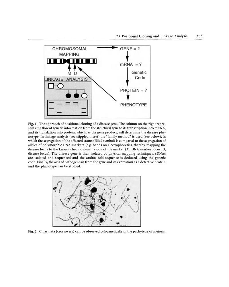
Fig. I. The approach of positional cloning of a disease gene. The column on the right represents the flow of genetic information from the structural gene to its transcription into mRNA,
and its translation into protein, which, as the gene product, will determine the disease phenotype. In linkage analysis (see stippled insert) the "family method" is used (see below), in
which the segregation of the affected status (filled symbol) is compared to the segregation of
alleles of polymorphic DNA markers (e.g. bands on electrophoresis), thereby mapping the
disease locus to the known chromosomal region of the marker (M, DNA marker locus; D,
disease locus). The disease gene is then isolated by physical mapping techniques. cDNAs
are isolated and sequenced and the amino acid sequence is deduced using the genetic
code. Finally, the axis of pathogenesis from the gene and its expression as a defective protein
and the phenotype can be studied.
Fig. 2. Chiasmata ( crossovers) can be observed cytogenetically in the pachytene of meiosis.
MAPPING
II I .,?'. Mll
M D
1=
23 Positional Cloning and Linkage Analysis
353
... GENE = ?
t
mRNA =?
I Genetic
+ Code
PROTEIN = ?
+
PHENOTYPE
Fig. I. The approach of positional cloning of a disease gene. The column on the right represents the flow of genetic information from the structural gene to its transcription into mRNA,
and its translation into protein, which, as the gene product, will determine the disease phenotype. In linkage analysis (see stippled insert) the "family method" is used (see below), in
which the segregation of the affected status (filled symbol) is compared to the segregation of
alleles of polymorphic DNA markers (e.g. bands on electrophoresis), thereby mapping the
disease locus to the known chromosomal region of the marker (M, DNA marker locus; D,
disease locus). The disease gene is then isolated by physical mapping techniques. cDNAs
are isolated and sequenced and the amino acid sequence is deduced using the genetic
code. Finally, the axis of pathogenesis from the gene and its expression as a defective protein
and the phenotype can be studied.
Fig. 2. Chiasmata ( crossovers) can be observed cytogenetically in the pachytene of meiosis.