

280
Assay of Some Common Hydrobiological Techniques
excess moisture is avoided by briefly opening the tap. The filter in the funnel
is rinsed with 1-2ml of prefiltered distilled water, and the set is ready for the
sample filtration. The portion of the fixed sample of 5-20 ml, after gentle
mixing, is first poured into the graduated cylinder to record volume (the use
of pipets for this procedure should be avoided). Then it is poured into the
funnel and slowly filtered under a vacuum of not more than 0.05 atm.
The speed of filtration is regulated with the tap. After the sample is passed,
the filter is immediately rinsed with 2-3 ml of distilled water, the tap is closed,
and the filter is soaked with primuline solution added to the funnel to stain
the algal cells. The time of staining should be adjusted empirically. Usually
it is between 5 and 15 min. Then the excess staining solution is filtered down
by opening the tap, the filter is rinsed again with 2-3 ml of distilled water, tap
is closed, and the still-moist Nucleopore filter is placed on the oiled area at
the slide. A drop of oil is placed on its surface, and it is covered with a cover
slide which is pressed until the whole surface of the filter is embedded into
the oil.
This preparation is ready for examination under the epifluorescence
microscope. The ocular of the microscope must be supplied with the counting
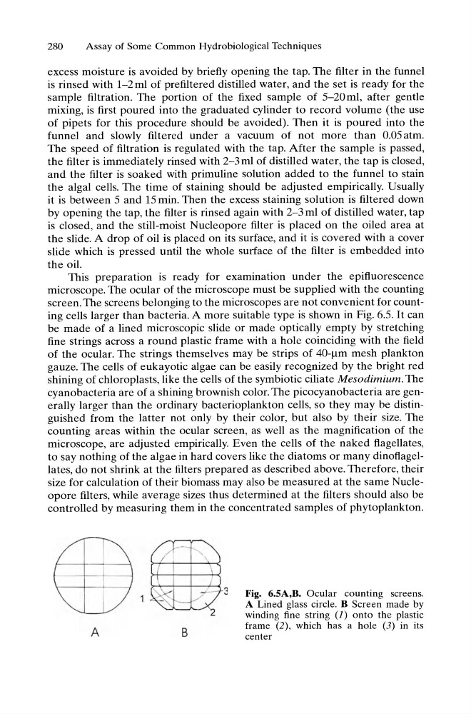
screen. The screens belonging to the microscopes are not convenient for counting cells larger than bacteria. A more suitable type is shown in Fig. 6.5. It can
be made of a lined microscopic slide or made optically empty by stretching
fine strings across a round plastic frame with a hole coinciding with the field
of the ocular. The strings themselves may be strips of 40-/lm mesh plankton
gauze. The cells of eukayotic algae can be easily recognized by the bright red
shining of chloroplasts, like the cells of the symbiotic ciliate Mesodimium. The
cyanobacteria are of a shining brownish color. The picocyanobacteria are generally larger than the ordinary bacterioplankton cells, so they may be distinguished from the latter not only by their color, but also by their size. The
counting areas within the ocular screen, as well as the magnification of the
microscope, are adjusted empirically. Even the cells of the naked flagellates,
to say nothing of the algae in hard covers like the diatoms or many dinoflagellates, do not shrink at the filters prepared as described above. Therefore, their
size for calculation of their biomass may also be measured at the same Nucleopore filters, while average sizes thus determined at the filters should also be
controlled by measuring them in the concentrated samples of phytoplankton.
A
B
3
Fig. 6.5A,B. Ocular counting screens.
A Lined glass circle. B Screen made by
winding fine string (1) onto the plastic
frame (2) , which has a hole (3) in its
center
Assay of Some Common Hydrobiological Techniques
excess moisture is avoided by briefly opening the tap. The filter in the funnel
is rinsed with 1-2ml of prefiltered distilled water, and the set is ready for the
sample filtration. The portion of the fixed sample of 5-20 ml, after gentle
mixing, is first poured into the graduated cylinder to record volume (the use
of pipets for this procedure should be avoided). Then it is poured into the
funnel and slowly filtered under a vacuum of not more than 0.05 atm.
The speed of filtration is regulated with the tap. After the sample is passed,
the filter is immediately rinsed with 2-3 ml of distilled water, the tap is closed,
and the filter is soaked with primuline solution added to the funnel to stain
the algal cells. The time of staining should be adjusted empirically. Usually
it is between 5 and 15 min. Then the excess staining solution is filtered down
by opening the tap, the filter is rinsed again with 2-3 ml of distilled water, tap
is closed, and the still-moist Nucleopore filter is placed on the oiled area at
the slide. A drop of oil is placed on its surface, and it is covered with a cover
slide which is pressed until the whole surface of the filter is embedded into
the oil.
This preparation is ready for examination under the epifluorescence
microscope. The ocular of the microscope must be supplied with the counting
screen. The screens belonging to the microscopes are not convenient for counting cells larger than bacteria. A more suitable type is shown in Fig. 6.5. It can
be made of a lined microscopic slide or made optically empty by stretching
fine strings across a round plastic frame with a hole coinciding with the field
of the ocular. The strings themselves may be strips of 40-/lm mesh plankton
gauze. The cells of eukayotic algae can be easily recognized by the bright red
shining of chloroplasts, like the cells of the symbiotic ciliate Mesodimium. The
cyanobacteria are of a shining brownish color. The picocyanobacteria are generally larger than the ordinary bacterioplankton cells, so they may be distinguished from the latter not only by their color, but also by their size. The
counting areas within the ocular screen, as well as the magnification of the
microscope, are adjusted empirically. Even the cells of the naked flagellates,
to say nothing of the algae in hard covers like the diatoms or many dinoflagellates, do not shrink at the filters prepared as described above. Therefore, their
size for calculation of their biomass may also be measured at the same Nucleopore filters, while average sizes thus determined at the filters should also be
controlled by measuring them in the concentrated samples of phytoplankton.
A
B
3
Fig. 6.5A,B. Ocular counting screens.
A Lined glass circle. B Screen made by
winding fine string (1) onto the plastic
frame (2) , which has a hole (3) in its
center