

Use of the Radiocarbon Method to Determine Primary Production
87
.~ vacuum
Ie
-....
- - - 'a
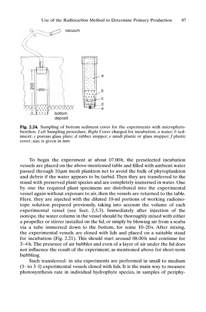
Fig. 2.24. Sampling of bottom sediment cores for the experiments with microphytobenthos. Left Sampling procedure. Right Corer charged for incubation. a water; b sediment; c porous glass plate; d rubber stopper; e small plastic or glass stopper; f plastic
corer; size is given in mm
To begin the experiment at about 07.00 h, the preselected incubation
vessels are placed on the above-mentioned table and filled with ambient water
passed through lOllm mesh plankton net to avoid the bulk of phytoplankton
and debris if the water appears to be turbid. Then they are transferred to the
stand with preserved plant species and are completely immersed in water. One
by one the required plant specimens are distributed into the experimental
vessel again without exposure to air, then the vessels are returned to the table.
Here, they are injected with the diluted lO-ml portions of working radioisotope solution prepared previously, taking into account the volume of each
experimental vessel (see Sect. 2.5.3). Immediately after injection of the
isotope, the water column in the vessel should be thoroughly mixed with either
a propeller or stirrer installed on the lid, or simply by blowing air from a scuba
via a tube immersed down to the bottom, for some 1O-20s. After mixing,
the experimental vessels are closed with lids and placed on a suitable stand
for incubation (Fig. 2.21). This should start around 08.00h and continue for
3-4 h. The presence of air bubbles and even of a layer of air under the lid does
not influence the result of the experiment, as mentioned above for short-term
bubbling.
Such transferred- in situ experiments are performed in small to medium
(1- to 3·1) experimental vessels closed with lids. It is the main way to measure
photosynthesis rate in individual hydrophyte species, in samples of periphy-
87
.~ vacuum
Ie
-....
- - - 'a
Fig. 2.24. Sampling of bottom sediment cores for the experiments with microphytobenthos. Left Sampling procedure. Right Corer charged for incubation. a water; b sediment; c porous glass plate; d rubber stopper; e small plastic or glass stopper; f plastic
corer; size is given in mm
To begin the experiment at about 07.00 h, the preselected incubation
vessels are placed on the above-mentioned table and filled with ambient water
passed through lOllm mesh plankton net to avoid the bulk of phytoplankton
and debris if the water appears to be turbid. Then they are transferred to the
stand with preserved plant species and are completely immersed in water. One
by one the required plant specimens are distributed into the experimental
vessel again without exposure to air, then the vessels are returned to the table.
Here, they are injected with the diluted lO-ml portions of working radioisotope solution prepared previously, taking into account the volume of each
experimental vessel (see Sect. 2.5.3). Immediately after injection of the
isotope, the water column in the vessel should be thoroughly mixed with either
a propeller or stirrer installed on the lid, or simply by blowing air from a scuba
via a tube immersed down to the bottom, for some 1O-20s. After mixing,
the experimental vessels are closed with lids and placed on a suitable stand
for incubation (Fig. 2.21). This should start around 08.00h and continue for
3-4 h. The presence of air bubbles and even of a layer of air under the lid does
not influence the result of the experiment, as mentioned above for short-term
bubbling.
Such transferred- in situ experiments are performed in small to medium
(1- to 3·1) experimental vessels closed with lids. It is the main way to measure
photosynthesis rate in individual hydrophyte species, in samples of periphy-