

39
Chapter two: Oxygen stores and diving
venous blood occurs in other marine mammals. However, as a mechanism to increase O 2
storage, and ultimately enhance breath-hold capacity, it would seem to be a highly beneficial mechanism.
Finally, in most mammals, the blood O 2 store cannot be completely depleted. Thus
to calculate the usable blood O 2 store, it is necessary to determine the minimum blood
O 2 level during dives. However, there have been few studies of blood O 2 store depletion
during diving. In marine mammals, the lowest arterial saturation is generally assumed to
be 20%. Forced submersion studies demonstrated that seals were tolerant of arterial Hb
saturation values down to about 20% (Elsner et al. 1970; Kerem and Elsner 1973). Tolerance
of low levels of O 2 in the blood is called hypoxemic tolerance. Northern elephant seals have
extreme hypoxemic tolerance with arterial Hb saturation often reaching values less than
5% (Meir et al. 2009). On the venous side, values near zero are often assumed as forced
submersion studies in seals have shown blood O 2 venous store can be completely depleted
(Kerem and Elsner 1973). In more recent studies on freely diving northern elephant seals
and California sea lions, venous Hb saturation values were near zero at times (Meir et al.
2009; McDonald and Ponganis 2013).
2.3.3 Measuring the muscle O 2 store
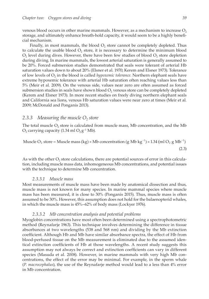
The total muscle O 2 store is calculated from muscle mass, Mb concentration, and the Mb
O 2 carrying capacity (1.34 ml O 2 g −1 Mb).
Muscle O store Muscle mass kg Mb concentration g Mb kg
1 34
2
1
=
´
´
-
( )
(
) . ( (
)
ml O g Mb
2
1
-
(2.3)
As with the other O 2 store calculations, there are potential sources of error in this calculation, including muscle mass data, inhomogeneous Mb concentrations, and potential issues
with the technique to determine Mb concentration.
2.3.3.1 Muscle mass
Most measurements of muscle mass have been made by anatomical dissection and thus,
muscle mass is not known for many species. In marine mammal species where muscle
mass has been measured, it is close to 30% (Ponganis 2015). Thus, muscle mass is often
assumed to be 30%. However, this assumption does not hold for the balaenopterid whales,
in which the muscle mass is 45%–62% of body mass (Lockyer 1976).
2.3.3.2 Mb concentration analysis and potential problems
Myoglobin concentrations have most often been determined using a spectrophotometric
method (Reynafarje 1963). This technique involves determining the difference in tissue
absorbances at two wavelengths (538 and 568 nm) and dividing by the Mb extinction
coefficient. Although Hb and Mb have similar absorbance spectra, the effect of Hb from
blood-perfused tissue on the Mb measurement is eliminated due to the assumed identical extinction coefficients of Hb at those wavelengths. A recent study suggests this
assumption may not always be correct and extinction coefficients can vary in different
species (Masuda et al. 2008). However, in marine mammals with very high Mb concentrations, the effect of the error may be minimal. For example, in the sperm whale
(P. macrocephalus), the use of the Reynafarje method would lead to a less than 4% error
in Mb concentration.
Chapter two: Oxygen stores and diving
venous blood occurs in other marine mammals. However, as a mechanism to increase O 2
storage, and ultimately enhance breath-hold capacity, it would seem to be a highly beneficial mechanism.
Finally, in most mammals, the blood O 2 store cannot be completely depleted. Thus
to calculate the usable blood O 2 store, it is necessary to determine the minimum blood
O 2 level during dives. However, there have been few studies of blood O 2 store depletion
during diving. In marine mammals, the lowest arterial saturation is generally assumed to
be 20%. Forced submersion studies demonstrated that seals were tolerant of arterial Hb
saturation values down to about 20% (Elsner et al. 1970; Kerem and Elsner 1973). Tolerance
of low levels of O 2 in the blood is called hypoxemic tolerance. Northern elephant seals have
extreme hypoxemic tolerance with arterial Hb saturation often reaching values less than
5% (Meir et al. 2009). On the venous side, values near zero are often assumed as forced
submersion studies in seals have shown blood O 2 venous store can be completely depleted
(Kerem and Elsner 1973). In more recent studies on freely diving northern elephant seals
and California sea lions, venous Hb saturation values were near zero at times (Meir et al.
2009; McDonald and Ponganis 2013).
2.3.3 Measuring the muscle O 2 store
The total muscle O 2 store is calculated from muscle mass, Mb concentration, and the Mb
O 2 carrying capacity (1.34 ml O 2 g −1 Mb).
Muscle O store Muscle mass kg Mb concentration g Mb kg
1 34
2
1
=
´
´
-
( )
(
) . ( (
)
ml O g Mb
2
1
-
(2.3)
As with the other O 2 store calculations, there are potential sources of error in this calculation, including muscle mass data, inhomogeneous Mb concentrations, and potential issues
with the technique to determine Mb concentration.
2.3.3.1 Muscle mass
Most measurements of muscle mass have been made by anatomical dissection and thus,
muscle mass is not known for many species. In marine mammal species where muscle
mass has been measured, it is close to 30% (Ponganis 2015). Thus, muscle mass is often
assumed to be 30%. However, this assumption does not hold for the balaenopterid whales,
in which the muscle mass is 45%–62% of body mass (Lockyer 1976).
2.3.3.2 Mb concentration analysis and potential problems
Myoglobin concentrations have most often been determined using a spectrophotometric
method (Reynafarje 1963). This technique involves determining the difference in tissue
absorbances at two wavelengths (538 and 568 nm) and dividing by the Mb extinction
coefficient. Although Hb and Mb have similar absorbance spectra, the effect of Hb from
blood-perfused tissue on the Mb measurement is eliminated due to the assumed identical extinction coefficients of Hb at those wavelengths. A recent study suggests this
assumption may not always be correct and extinction coefficients can vary in different
species (Masuda et al. 2008). However, in marine mammals with very high Mb concentrations, the effect of the error may be minimal. For example, in the sperm whale
(P. macrocephalus), the use of the Reynafarje method would lead to a less than 4% error
in Mb concentration.