

38
Marine Mammal Physiology: Requisites for Ocean Living
decrease during a dive from 95% to 20%, and that the venous O 2 , with an initial venous
content of 5 ml O 2 dl −1 less than the arterial value, can be completely depleted. This has
been estimated in most cases, but has been calculated as the difference between the initial
O 2 value at the beginning of the dive and the lowest O 2 value obtained in any dive (or the
difference between the beginning and end values for SaO 2 and SvO 2 ) (Lenfant et al. 1970;
Kooyman 1989). See Section 2.3.2.3 for further review.
2.3.2.1 Blood volume
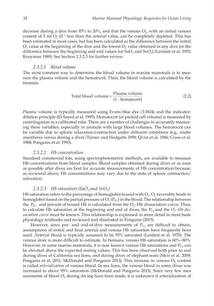
The most common way to determine the blood volume in marine mammals is to measure the plasma volume and the hematocrit. Then, the blood volume is calculated by the
formula:
Total blood volume
Plasma volume
1 hematocrit
= -
(
)
(2.2)
Plasma volume is typically measured using Evans blue dye (T-1824) and the indicatordilution principle (El-Sayed et al. 1995). Hematocrit (or packed cell volume) is measured by
centrifugation in a calibrated tube. There are a number of challenges in accurately measuring these variables, especially in animals with large blood volumes. The hematocrit can
be variable due to splenic relaxation/contraction under different conditions (e.g., under
anesthesia versus during a dive) (Turner and Hodgetts 1959; Qvist et al. 1986; Cross et al.
1988; Ponganis et al. 1993).
2.3.2.2 Hb concentration
Standard commercial kits, using spectrophotometric methods, are available to measure
Hb concentrations from blood samples. Blood samples obtained during dives or as soon
as possible after dives are best for accurate measurements of Hb concentration because,
as reviewed above, Hb concentrations may vary due to the state of splenic contraction/
relaxation.
2.3.2.3 Hb saturation (SaO 2 and SvO 2 )
Hb saturation refers to the percentage of hemoglobin bound with O 2 . O 2 reversibly binds to
hemoglobin based on the partial pressure of O 2 (P O2 ) in the blood. The relationship between
the P O2 and percent of bound Hb is calculated from the O 2 –Hb dissociation curve. Thus,
to calculate Hb saturation at the beginning and end of dives, the P O2 and the O 2 –Hb dissociation curve must be known. This relationship is explained in more detail in most basic
physiology textbooks and reviewed and illustrated in Ponganis (2015).
However, since pre- and end-of-dive measurements of P O2 are difficult to obtain,
assumptions of initial and final arterial and venous Hb saturation have frequently been
used. Arterial blood is typically assumed to be 95% saturated (Lenfant et al. 1970). The
venous store is more difficult to estimate. In humans, venous Hb saturation is 60%–80%.
However, in some marine mammals, it is now known venous Hb saturations and P O2 can
be elevated above the expected resting values. This has been observed both prior to and
during dives of California sea lions, and during dives of elephant seals (Meir et al. 2009;
Ponganis et al. 2011; McDonald and Ponganis 2013). This increase in venous O 2 content
is called arterialization of venous blood. In sea lions, the venous blood in some dives was
increased to above 95% saturation (McDonald and Ponganis 2013). Since very few mea
surements of blood O 2 during diving have been made, it is unknown if arterialization of
Marine Mammal Physiology: Requisites for Ocean Living
decrease during a dive from 95% to 20%, and that the venous O 2 , with an initial venous
content of 5 ml O 2 dl −1 less than the arterial value, can be completely depleted. This has
been estimated in most cases, but has been calculated as the difference between the initial
O 2 value at the beginning of the dive and the lowest O 2 value obtained in any dive (or the
difference between the beginning and end values for SaO 2 and SvO 2 ) (Lenfant et al. 1970;
Kooyman 1989). See Section 2.3.2.3 for further review.
2.3.2.1 Blood volume
The most common way to determine the blood volume in marine mammals is to measure the plasma volume and the hematocrit. Then, the blood volume is calculated by the
formula:
Total blood volume
Plasma volume
1 hematocrit
= -
(
)
(2.2)
Plasma volume is typically measured using Evans blue dye (T-1824) and the indicatordilution principle (El-Sayed et al. 1995). Hematocrit (or packed cell volume) is measured by
centrifugation in a calibrated tube. There are a number of challenges in accurately measuring these variables, especially in animals with large blood volumes. The hematocrit can
be variable due to splenic relaxation/contraction under different conditions (e.g., under
anesthesia versus during a dive) (Turner and Hodgetts 1959; Qvist et al. 1986; Cross et al.
1988; Ponganis et al. 1993).
2.3.2.2 Hb concentration
Standard commercial kits, using spectrophotometric methods, are available to measure
Hb concentrations from blood samples. Blood samples obtained during dives or as soon
as possible after dives are best for accurate measurements of Hb concentration because,
as reviewed above, Hb concentrations may vary due to the state of splenic contraction/
relaxation.
2.3.2.3 Hb saturation (SaO 2 and SvO 2 )
Hb saturation refers to the percentage of hemoglobin bound with O 2 . O 2 reversibly binds to
hemoglobin based on the partial pressure of O 2 (P O2 ) in the blood. The relationship between
the P O2 and percent of bound Hb is calculated from the O 2 –Hb dissociation curve. Thus,
to calculate Hb saturation at the beginning and end of dives, the P O2 and the O 2 –Hb dissociation curve must be known. This relationship is explained in more detail in most basic
physiology textbooks and reviewed and illustrated in Ponganis (2015).
However, since pre- and end-of-dive measurements of P O2 are difficult to obtain,
assumptions of initial and final arterial and venous Hb saturation have frequently been
used. Arterial blood is typically assumed to be 95% saturated (Lenfant et al. 1970). The
venous store is more difficult to estimate. In humans, venous Hb saturation is 60%–80%.
However, in some marine mammals, it is now known venous Hb saturations and P O2 can
be elevated above the expected resting values. This has been observed both prior to and
during dives of California sea lions, and during dives of elephant seals (Meir et al. 2009;
Ponganis et al. 2011; McDonald and Ponganis 2013). This increase in venous O 2 content
is called arterialization of venous blood. In sea lions, the venous blood in some dives was
increased to above 95% saturation (McDonald and Ponganis 2013). Since very few mea
surements of blood O 2 during diving have been made, it is unknown if arterialization of