

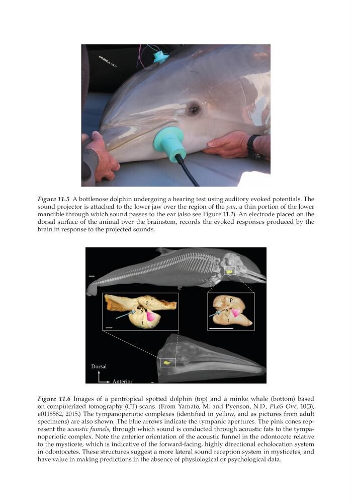
Figure 11.5 A bottlenose dolphin undergoing a hearing test using auditory evoked potentials. The
sound projector is attached to the lower jaw over the region of the pan, a thin portion of the lower
mandible through which sound passes to the ear (also see Figure 11.2). An electrode placed on the
dorsal surface of the animal over the brainstem, records the evoked responses produced by the
brain in response to the projected sounds.
Dorsal
Anterior
p
s
t
p
s
t
Figure 11.6 Images of a pantropical spotted dolphin (top) and a minke whale (bottom) based
on computerized tomography (CT) scans. (From Yamato, M. and Pyenson, N.D., PLoS One, 10(3),
e0118582, 2015.) The tympanoperiotic complexes (identified in yellow, and as pictures from adult
specimens) are also shown. The blue arrows indicate the tympanic apertures. The pink cones represent the acoustic funnels, through which sound is conducted through acoustic fats to the tympanoperiotic complex. Note the anterior orientation of the acoustic funnel in the odontocete relative
to the mysticete, which is indicative of the forward-facing, highly directional echolocation system
in odontocetes. These structures suggest a more lateral sound reception system in mysticetes, and
have value in making predictions in the absence of physiological or psychological data.
sound projector is attached to the lower jaw over the region of the pan, a thin portion of the lower
mandible through which sound passes to the ear (also see Figure 11.2). An electrode placed on the
dorsal surface of the animal over the brainstem, records the evoked responses produced by the
brain in response to the projected sounds.
Dorsal
Anterior
p
s
t
p
s
t
Figure 11.6 Images of a pantropical spotted dolphin (top) and a minke whale (bottom) based
on computerized tomography (CT) scans. (From Yamato, M. and Pyenson, N.D., PLoS One, 10(3),
e0118582, 2015.) The tympanoperiotic complexes (identified in yellow, and as pictures from adult
specimens) are also shown. The blue arrows indicate the tympanic apertures. The pink cones represent the acoustic funnels, through which sound is conducted through acoustic fats to the tympanoperiotic complex. Note the anterior orientation of the acoustic funnel in the odontocete relative
to the mysticete, which is indicative of the forward-facing, highly directional echolocation system
in odontocetes. These structures suggest a more lateral sound reception system in mysticetes, and
have value in making predictions in the absence of physiological or psychological data.