

262
Marine Mammal Physiology: Requisites for Ocean Living
hearing ranges has been derived from anatomical models (Houser et al. 2001; Parks et al.
2007; Tubelli et al. 2012). Unfortunately, without validation of the models, there remains
much uncertainty as to their predictive capability. The use of AEP methods, previously
mentioned, may provide one of the better opportunities for measuring hearing in mysticetes. However, this, too, is fraught with challenges. Evoked potential methods work well with
odontocetes because they have a large auditory nerve, smooth skin, thin skull, thin blubber
layers (for many species), a large brain:body mass ratio, and highly refined auditory processing. On the other hand, mysticetes have large and thick skulls, large blubber layers that
attenuate electrical signals recorded at the body surface (i.e., blubber is a poor conductor of
electricity), a largely unfavorable brain:body mass ratio, and a less derived auditory system.
These factors either contribute to small amplitude AEPs or make the recording of the AEPs
difficult, at least by non-invasive methods. Nevertheless, the first empirical measurements
of hearing in a mysticete whale may likely have to be made through electrophysiological
methods but may have to be performed on the smallest species (e.g., minke whale) or calves
of relatively small species (e.g., gray whale calves) for early successes.
A related controversy exists with our knowledge of hearing in odontocetes. Although
research primarily conducted on bottlenose dolphins has provided considerable information about the peripheral auditory pathway and how sound enters the dolphin head,
it remains unclear as to how acoustic energy is conducted to the cochlea. Is the stapes
vibrated by the malleus and incus such that it manipulates the oval window, as occurs
Dorsal
Anterior
p
s
t
p
s
t
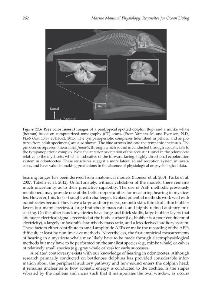
Figure 11.6 (See color insert.) Images of a pantropical spotted dolphin (top) and a minke whale
(bottom) based on computerized tomography (CT) scans. (From Yamato, M. and Pyenson, N.D.,
PLoS One, 10(3), e0118582, 2015.) The tympanoperiotic complexes (identified in yellow, and as pictures from adult specimens) are also shown. The blue arrows indicate the tympanic apertures. The
pink cones represent the acoustic funnels, through which sound is conducted through acoustic fats to
the tympanoperiotic complex. Note the anterior orientation of the acoustic funnel in the odontocete
relative to the mysticete, which is indicative of the forward-facing, highly directional echolocation
system in odontocetes. These structures suggest a more lateral sound reception system in mysticetes, and have value in making predictions in the absence of physiological or psychological data.
Marine Mammal Physiology: Requisites for Ocean Living
hearing ranges has been derived from anatomical models (Houser et al. 2001; Parks et al.
2007; Tubelli et al. 2012). Unfortunately, without validation of the models, there remains
much uncertainty as to their predictive capability. The use of AEP methods, previously
mentioned, may provide one of the better opportunities for measuring hearing in mysticetes. However, this, too, is fraught with challenges. Evoked potential methods work well with
odontocetes because they have a large auditory nerve, smooth skin, thin skull, thin blubber
layers (for many species), a large brain:body mass ratio, and highly refined auditory processing. On the other hand, mysticetes have large and thick skulls, large blubber layers that
attenuate electrical signals recorded at the body surface (i.e., blubber is a poor conductor of
electricity), a largely unfavorable brain:body mass ratio, and a less derived auditory system.
These factors either contribute to small amplitude AEPs or make the recording of the AEPs
difficult, at least by non-invasive methods. Nevertheless, the first empirical measurements
of hearing in a mysticete whale may likely have to be made through electrophysiological
methods but may have to be performed on the smallest species (e.g., minke whale) or calves
of relatively small species (e.g., gray whale calves) for early successes.
A related controversy exists with our knowledge of hearing in odontocetes. Although
research primarily conducted on bottlenose dolphins has provided considerable information about the peripheral auditory pathway and how sound enters the dolphin head,
it remains unclear as to how acoustic energy is conducted to the cochlea. Is the stapes
vibrated by the malleus and incus such that it manipulates the oval window, as occurs
Dorsal
Anterior
p
s
t
p
s
t
Figure 11.6 (See color insert.) Images of a pantropical spotted dolphin (top) and a minke whale
(bottom) based on computerized tomography (CT) scans. (From Yamato, M. and Pyenson, N.D.,
PLoS One, 10(3), e0118582, 2015.) The tympanoperiotic complexes (identified in yellow, and as pictures from adult specimens) are also shown. The blue arrows indicate the tympanic apertures. The
pink cones represent the acoustic funnels, through which sound is conducted through acoustic fats to
the tympanoperiotic complex. Note the anterior orientation of the acoustic funnel in the odontocete
relative to the mysticete, which is indicative of the forward-facing, highly directional echolocation
system in odontocetes. These structures suggest a more lateral sound reception system in mysticetes, and have value in making predictions in the absence of physiological or psychological data.