

247
Chapter eleven: Acoustics
The cetaceans, which consist of the toothed and baleen whales, are undoubtedly the
most derived of the marine mammals when it comes to the auditory system. Having an
evolutionary history traced through ungulate ancestors, the Cetacea demonstrate adaptations to the auditory system and vestibular systems that are unique relative to other
mammalian species. However, within the Cetacea there are dramatic differences between
the suborders Odontoceti (toothed whales) and Mysticeti (baleen whales), with the former
being the more derived of the two suborders.
The overall morphology of the cetacean head has resulted from selective pressures
related to underwater movement efficiency (e.g., reduction in drag) and surface respiration. Cetaceans typically demonstrate a streamlined, fusiform shape (see Chapter 1)
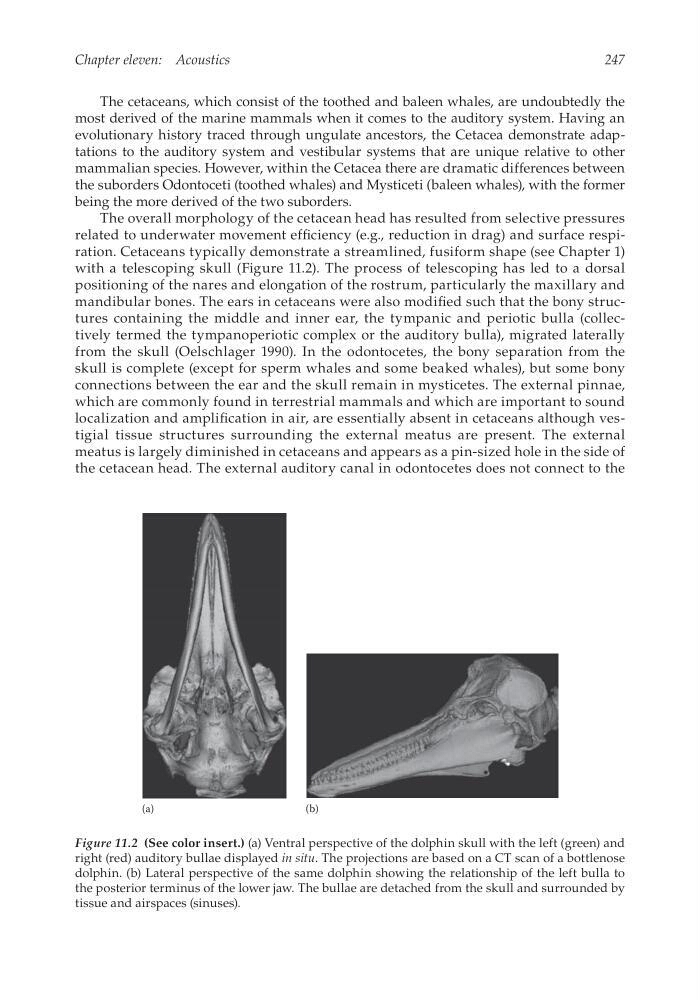
with a telescoping skull (Figure 11.2). The process of telescoping has led to a dorsal
positioning of the nares and elongation of the rostrum, particularly the maxillary and
mandibular bones. The ears in cetaceans were also modified such that the bony structures containing the middle and inner ear, the tympanic and periotic bulla (collectively termed the tympanoperiotic complex or the auditory bulla), migrated laterally
from the skull (Oelschlager 1990). In the odontocetes, the bony separation from the
skull is complete (except for sperm whales and some beaked whales), but some bony
connections between the ear and the skull remain in mysticetes. The external pinnae,
which are commonly found in terrestrial mammals and which are important to sound
localization and amplification in air, are essentially absent in cetaceans although vestigial tissue structures surrounding the external meatus are present. The external
meatus is largely diminished in cetaceans and appears as a pin-sized hole in the side of
the cetacean head. The external auditory canal in odontocetes does not connect to the
(a)
(b)
Figure 11.2 (See color insert.) (a) Ventral perspective of the dolphin skull with the left (green) and
right (red) auditory bullae displayed in situ. The projections are based on a CT scan of a bottlenose
dolphin. (b) Lateral perspective of the same dolphin showing the relationship of the left bulla to
the posterior terminus of the lower jaw. The bullae are detached from the skull and surrounded by
t issue and airspaces (sinuses).
Chapter eleven: Acoustics
The cetaceans, which consist of the toothed and baleen whales, are undoubtedly the
most derived of the marine mammals when it comes to the auditory system. Having an
evolutionary history traced through ungulate ancestors, the Cetacea demonstrate adaptations to the auditory system and vestibular systems that are unique relative to other
mammalian species. However, within the Cetacea there are dramatic differences between
the suborders Odontoceti (toothed whales) and Mysticeti (baleen whales), with the former
being the more derived of the two suborders.
The overall morphology of the cetacean head has resulted from selective pressures
related to underwater movement efficiency (e.g., reduction in drag) and surface respiration. Cetaceans typically demonstrate a streamlined, fusiform shape (see Chapter 1)
with a telescoping skull (Figure 11.2). The process of telescoping has led to a dorsal
positioning of the nares and elongation of the rostrum, particularly the maxillary and
mandibular bones. The ears in cetaceans were also modified such that the bony structures containing the middle and inner ear, the tympanic and periotic bulla (collectively termed the tympanoperiotic complex or the auditory bulla), migrated laterally
from the skull (Oelschlager 1990). In the odontocetes, the bony separation from the
skull is complete (except for sperm whales and some beaked whales), but some bony
connections between the ear and the skull remain in mysticetes. The external pinnae,
which are commonly found in terrestrial mammals and which are important to sound
localization and amplification in air, are essentially absent in cetaceans although vestigial tissue structures surrounding the external meatus are present. The external
meatus is largely diminished in cetaceans and appears as a pin-sized hole in the side of
the cetacean head. The external auditory canal in odontocetes does not connect to the
(a)
(b)
Figure 11.2 (See color insert.) (a) Ventral perspective of the dolphin skull with the left (green) and
right (red) auditory bullae displayed in situ. The projections are based on a CT scan of a bottlenose
dolphin. (b) Lateral perspective of the same dolphin showing the relationship of the left bulla to
the posterior terminus of the lower jaw. The bullae are detached from the skull and surrounded by
t issue and airspaces (sinuses).