

Ontogeny of Marine Turtle Gonads
209
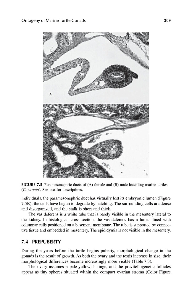
individuals, the paramesonephric duct has virtually lost its embryonic lumen (Figure
7.5B); the cells have begun to degrade by hatching. The surrounding cells are dense
and disorganized, and the stalk is short and thick.
The vas deferens is a white tube that is barely visible in the mesentery lateral to
the kidney. In histological cross section, the vas deferens has a lumen lined with
columnar cells positioned on a basement membrane. The tube is supported by connective tissue and embedded in mesentery. The epididymis is not visible in the mesentery.
7.4 PREPUBERTY
During the years before the turtle begins puberty, morphological change in the
gonads is the result of growth. As both the ovary and the testis increase in size, their
morphological differences become increasingly more visible (Table 7.3).
The ovary assumes a pale-yellowish tinge, and the previtellogenetic follicles
appear as tiny spheres situated within the compact ovarian stroma (Color Figure
FIGURE 7.5 Paramesonephric ducts of (A) female and (B) male hatchling marine turtles
( C. caretta ). See text for descriptions.
1123 book.book Page 209 Monday, November 11, 2002 11:11 AM
209
individuals, the paramesonephric duct has virtually lost its embryonic lumen (Figure
7.5B); the cells have begun to degrade by hatching. The surrounding cells are dense
and disorganized, and the stalk is short and thick.
The vas deferens is a white tube that is barely visible in the mesentery lateral to
the kidney. In histological cross section, the vas deferens has a lumen lined with
columnar cells positioned on a basement membrane. The tube is supported by connective tissue and embedded in mesentery. The epididymis is not visible in the mesentery.
7.4 PREPUBERTY
During the years before the turtle begins puberty, morphological change in the
gonads is the result of growth. As both the ovary and the testis increase in size, their
morphological differences become increasingly more visible (Table 7.3).
The ovary assumes a pale-yellowish tinge, and the previtellogenetic follicles
appear as tiny spheres situated within the compact ovarian stroma (Color Figure
FIGURE 7.5 Paramesonephric ducts of (A) female and (B) male hatchling marine turtles
( C. caretta ). See text for descriptions.
1123 book.book Page 209 Monday, November 11, 2002 11:11 AM