

208
The Biology of Sea Turtles, Vol. II
7.3.3 G ENITAL D UCTS
The paramesonephric (müllerian duct, oviduct) in the female is white and straight,
and can be seen traversing the posterior of the kidney. In histological cross section,
the lumen is open and lined with columnar cells that sit on a distinct basement
membrane (Figure 7.5A). The surrounding tissues are penetrated by blood vessels.
The suspending mesentery is long and thin. Histologically in the male, in most
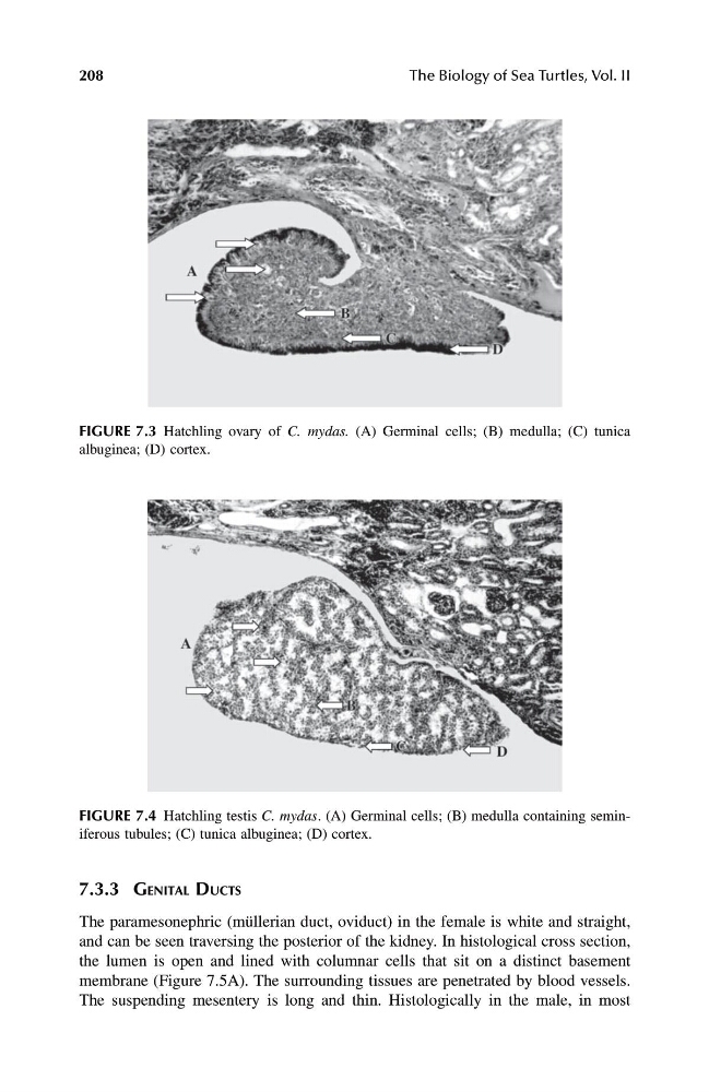
FIGURE 7.3 Hatchling ovary of C. mydas. (A) Germinal cells; (B) medulla; (C) tunica
albuginea; (D) cortex.
FIGURE 7.4 Hatchling testis C. mydas . (A) Germinal cells; (B) medulla containing seminiferous tubules; (C) tunica albuginea; (D) cortex.
1123 book.book Page 208 Monday, November 11, 2002 11:11 AM
The Biology of Sea Turtles, Vol. II
7.3.3 G ENITAL D UCTS
The paramesonephric (müllerian duct, oviduct) in the female is white and straight,
and can be seen traversing the posterior of the kidney. In histological cross section,
the lumen is open and lined with columnar cells that sit on a distinct basement
membrane (Figure 7.5A). The surrounding tissues are penetrated by blood vessels.
The suspending mesentery is long and thin. Histologically in the male, in most
FIGURE 7.3 Hatchling ovary of C. mydas. (A) Germinal cells; (B) medulla; (C) tunica
albuginea; (D) cortex.
FIGURE 7.4 Hatchling testis C. mydas . (A) Germinal cells; (B) medulla containing seminiferous tubules; (C) tunica albuginea; (D) cortex.
1123 book.book Page 208 Monday, November 11, 2002 11:11 AM