

The External, Musculoskeletal and Neuro-Anatomy of Sea Turtles
51
the palate’s midline, anterior to the vomer. In both Lepidochelys species, the two
maxillary bones are separated by the vomer, which extends anteriorly to articulate
with the premaxillary bones (Figure 2.11). The palate of L. kempii has longitudinal
alveolar ridges, whereas that of L. olivacea lacks alveolar ridges. The pterygoid
bones of the olive ridley are proportionately wider and the pterygoid processes are
more pronounced than in the Kemp’s ridley.
The hawksbill head is long and narrow (Figure 2.1B) in all but very young
turtles; its length is approximately equal to twice the width (measured at the skull’s
widest part). Hawksbill skulls have deep parietal notches, and the snout tapers to a
point. The secondary palate is well developed. As a result the internal nares are
situated slightly more posteriorly than in other cheloniids.
The skull of sea turtles articulates with the cervical vertebrae via a single, trilobed
occipital condyle formed by three occipital bones (Figure 2.9B).
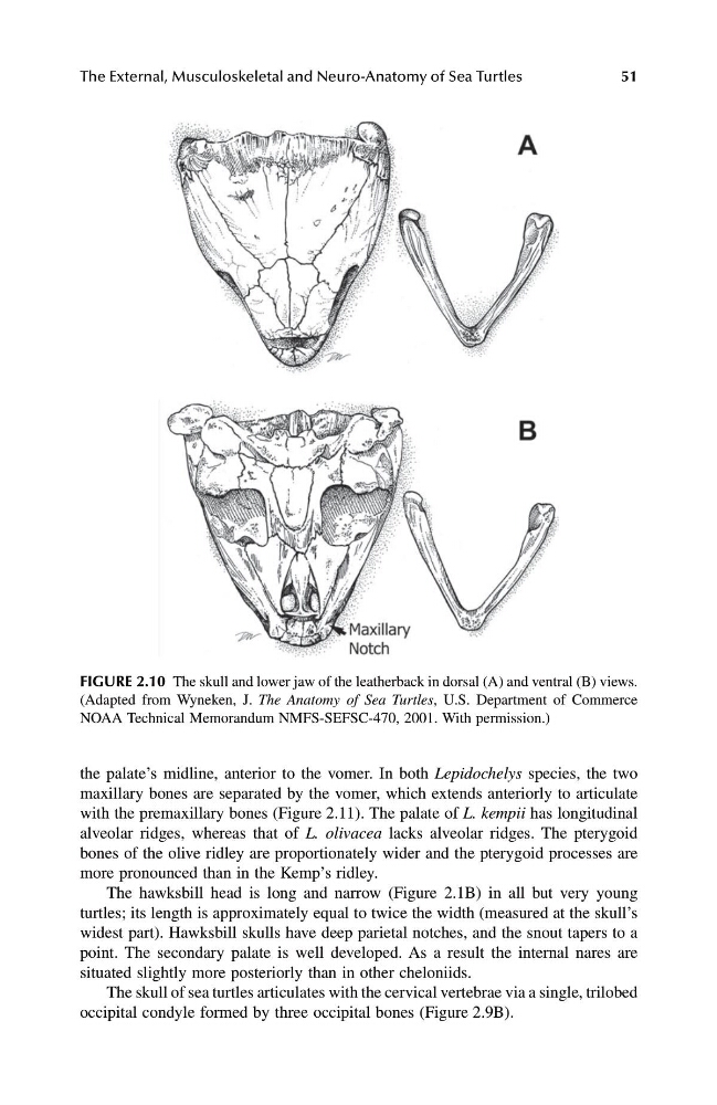
FIGURE 2.10 The skull and lower jaw of the leatherback in dorsal (A) and ventral (B) views.
(Adapted from Wyneken, J. The Anatomy of Sea Turtles, U.S. Department of Commerce
NOAA Technical Memorandum NMFS-SEFSC-470, 2001. With permission.)
1123_C02.fm Page 51 Friday, November 15, 2002 11:16 AM
51
the palate’s midline, anterior to the vomer. In both Lepidochelys species, the two
maxillary bones are separated by the vomer, which extends anteriorly to articulate
with the premaxillary bones (Figure 2.11). The palate of L. kempii has longitudinal
alveolar ridges, whereas that of L. olivacea lacks alveolar ridges. The pterygoid
bones of the olive ridley are proportionately wider and the pterygoid processes are
more pronounced than in the Kemp’s ridley.
The hawksbill head is long and narrow (Figure 2.1B) in all but very young
turtles; its length is approximately equal to twice the width (measured at the skull’s
widest part). Hawksbill skulls have deep parietal notches, and the snout tapers to a
point. The secondary palate is well developed. As a result the internal nares are
situated slightly more posteriorly than in other cheloniids.
The skull of sea turtles articulates with the cervical vertebrae via a single, trilobed
occipital condyle formed by three occipital bones (Figure 2.9B).
FIGURE 2.10 The skull and lower jaw of the leatherback in dorsal (A) and ventral (B) views.
(Adapted from Wyneken, J. The Anatomy of Sea Turtles, U.S. Department of Commerce
NOAA Technical Memorandum NMFS-SEFSC-470, 2001. With permission.)
1123_C02.fm Page 51 Friday, November 15, 2002 11:16 AM