

The External, Musculoskeletal and Neuro-Anatomy of Sea Turtles
49
2.3.1 THE SKULL
The skull is formed by an inner braincase (the neurocranium) housing the brain,
and an outer set of bones (the splanchnocranium). The braincase is found along the
midline, internal to the skull roof, snout, and jaws of the splanchnocranium. The
splanchnocranium houses the sense organs and provides the attachment sites for jaw,
throat, and neck muscles (Hyman and Wake, 1979; Kardong, 2002). The eyes each
contain a ring of bones (scleral ossicles) that, in life, are found deep to the iris. Skull
shape plus the form and the patterns of bones on the roof of the mouth are diagnostic
for species identification (Pritchard, 1979).
The jaws (mandibles) of sea turtles are composed of several articulating bones
and Meckel’s cartilage, within the lower mandible (Figure 2.9). Rhamphotheci cover
the maxillary, premaxillary, and vomer bones of the upper jaw, and the dentary bones
of the lower jaw in cheloniids.
Leatherback skulls are wide posteriorly, are rounded anteriorly, and possess large
orbits; there are no parietal notches. The skull is composed of loosely articulated
bones. There is no prominent supraoccipital process (Figure 2.10A). The jaws are
characterized by the presence of a notch in each maxillary bone (Figure 2.10B). The
lower jaw has a large cartilaginous portion, medial to the dentary. Anteriorly, the
two mandibles articulate at the midline to form a sharp point.
Cheloniid skulls are formed by tightly articulating bones. There is a partial
secondary palate present (Figure 2.11). Overall skull form and the details of the
palate differ among species. In C. mydas, the skull is rounded with a short snout
and shallow parietal notches. The palate, between the margins of the upper jaw and
the internal nares, has a pair of ridges that run parallel to the outer edge of the jaw.
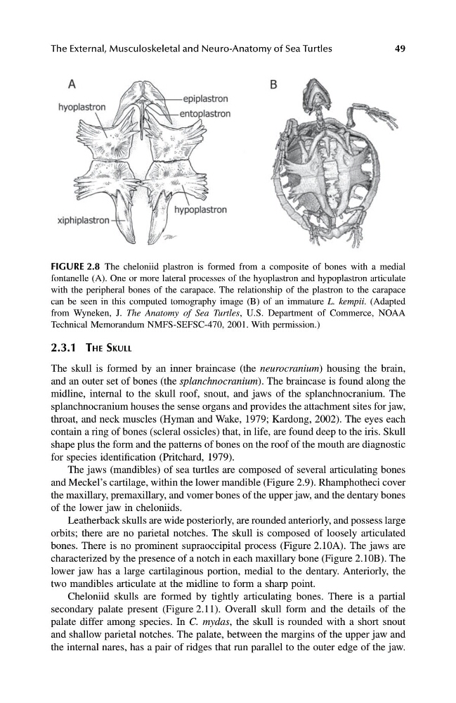
FIGURE 2.8 The cheloniid plastron is formed from a composite of bones with a medial
fontanelle (A). One or more lateral processes of the hyoplastron and hypoplastron articulate
with the peripheral bones of the carapace. The relationship of the plastron to the carapace
can be seen in this computed tomography image (B) of an immature L. kempii. (Adapted
from Wyneken, J. The Anatomy of Sea Turtles, U.S. Department of Commerce, NOAA
Technical Memorandum NMFS-SEFSC-470, 2001. With permission.)
1123_C02.fm Page 49 Friday, November 15, 2002 11:16 AM
49
2.3.1 THE SKULL
The skull is formed by an inner braincase (the neurocranium) housing the brain,
and an outer set of bones (the splanchnocranium). The braincase is found along the
midline, internal to the skull roof, snout, and jaws of the splanchnocranium. The
splanchnocranium houses the sense organs and provides the attachment sites for jaw,
throat, and neck muscles (Hyman and Wake, 1979; Kardong, 2002). The eyes each
contain a ring of bones (scleral ossicles) that, in life, are found deep to the iris. Skull
shape plus the form and the patterns of bones on the roof of the mouth are diagnostic
for species identification (Pritchard, 1979).
The jaws (mandibles) of sea turtles are composed of several articulating bones
and Meckel’s cartilage, within the lower mandible (Figure 2.9). Rhamphotheci cover
the maxillary, premaxillary, and vomer bones of the upper jaw, and the dentary bones
of the lower jaw in cheloniids.
Leatherback skulls are wide posteriorly, are rounded anteriorly, and possess large
orbits; there are no parietal notches. The skull is composed of loosely articulated
bones. There is no prominent supraoccipital process (Figure 2.10A). The jaws are
characterized by the presence of a notch in each maxillary bone (Figure 2.10B). The
lower jaw has a large cartilaginous portion, medial to the dentary. Anteriorly, the
two mandibles articulate at the midline to form a sharp point.
Cheloniid skulls are formed by tightly articulating bones. There is a partial
secondary palate present (Figure 2.11). Overall skull form and the details of the
palate differ among species. In C. mydas, the skull is rounded with a short snout
and shallow parietal notches. The palate, between the margins of the upper jaw and
the internal nares, has a pair of ridges that run parallel to the outer edge of the jaw.
FIGURE 2.8 The cheloniid plastron is formed from a composite of bones with a medial
fontanelle (A). One or more lateral processes of the hyoplastron and hypoplastron articulate
with the peripheral bones of the carapace. The relationship of the plastron to the carapace
can be seen in this computed tomography image (B) of an immature L. kempii. (Adapted
from Wyneken, J. The Anatomy of Sea Turtles, U.S. Department of Commerce, NOAA
Technical Memorandum NMFS-SEFSC-470, 2001. With permission.)
1123_C02.fm Page 49 Friday, November 15, 2002 11:16 AM