

Pathogenic Free-Living A moebae and Recreational Waters
91
of 3-ml samples for cell sheet plaquing to enumerate the pathogenic Naegleria. Decimal
dilutions were prepared from the samples and used in both plaquing when plaque counts
were expected to be high. The duplicate plaqued plates were incubated at room
temperature and the plaqued cell sheets in bottles were incubated at 35°C and were
examined for appearance of plaques daily for 2 weeks. To free the samples of bacteria for
plaquing on cell sheets, each sample was washed and centrifuged 4 times with dilution
water containing 50 Mg/ml of tetracycline and streptomycin.
The averages of the duplicate plaque counts during a 6-week study are plotted
graphically in Fig. 1. The growth curves of the strains of Naegleria in the figure show
clearly that while the wild strain was growing in a manner similar to that ascertained in
plate cultures (Chang, 1958), the pathogenic strain showed a steady decrease in
population and reached almost its extinction in a 6-week period.
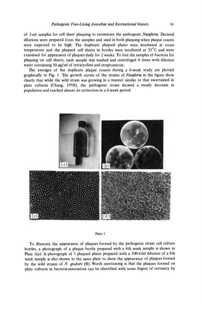
Plate 1
To illustrate the appearance of plaques formed by the pathogenic strain cell culture
bottles, a photograph of a plaque bottle prepared with a 4th week sample is shown in
Plate 1(a). A photograph of 3 plaqued plates prepared with a 100-fold dilution of a 4th
week sample is also shown in the same plate to show the appearance of plaques formed
by the wild strains of N gruben (B). Worth mentioning is that the plaques formed on
plate cultures in bacteria-association can be identified with some degree of certainty by
91
of 3-ml samples for cell sheet plaquing to enumerate the pathogenic Naegleria. Decimal
dilutions were prepared from the samples and used in both plaquing when plaque counts
were expected to be high. The duplicate plaqued plates were incubated at room
temperature and the plaqued cell sheets in bottles were incubated at 35°C and were
examined for appearance of plaques daily for 2 weeks. To free the samples of bacteria for
plaquing on cell sheets, each sample was washed and centrifuged 4 times with dilution
water containing 50 Mg/ml of tetracycline and streptomycin.
The averages of the duplicate plaque counts during a 6-week study are plotted
graphically in Fig. 1. The growth curves of the strains of Naegleria in the figure show
clearly that while the wild strain was growing in a manner similar to that ascertained in
plate cultures (Chang, 1958), the pathogenic strain showed a steady decrease in
population and reached almost its extinction in a 6-week period.
Plate 1
To illustrate the appearance of plaques formed by the pathogenic strain cell culture
bottles, a photograph of a plaque bottle prepared with a 4th week sample is shown in
Plate 1(a). A photograph of 3 plaqued plates prepared with a 100-fold dilution of a 4th
week sample is also shown in the same plate to show the appearance of plaques formed
by the wild strains of N gruben (B). Worth mentioning is that the plaques formed on
plate cultures in bacteria-association can be identified with some degree of certainty by