

The optional expression of genes may be visualized through recently developed
technology known as a DNA microarray (Fig. 3.2a). A DNA microarray is a glass
slide that localizes thousands or more types of single-stranded DNA fragments
arranged on an array (lattice). Each DNA fragment is obtained from a single gene,
so that a single microarray contains thousands of genes’ worth of DNA.
To use a microarray, researchers first assemble all mRNA expressed in a particular cell ①. As the mRNA is mixed with the viral enzyme reverse transcriptase,
this results in ② synthesis of complementary DNA for each mRNA. This complementary DNA (cDNA) is synthesized with nucleotides marked with fluorescent
substances. Small quantities of the fluorescent-marked cDNA mixture are then ③
mixed with thousands of kinds of single-stranded DNA in the microarray. If a
molecule in the cDNA mixture complements a piece of DNA at a particular position
in the microarray, that cDNA molecule binds with its corresponding DNA fragment
and remains fixed at that location. Once the unbound cDNA molecules are washed
off ④, the cDNA molecules remaining on the microarray become fluorescent
(Fig. 3.2b). Observing the patterns in the fluorescent areas allows researchers to
determine what genes have been turned on and off in the cell (Schena et al. 1995).
Microarray experiments have allowed researchers to observe differences in gene
expression between different tissues and tissues in humans with different health
conditions. It is a powerful new technology that has opened new horizons for gene
regulation research.
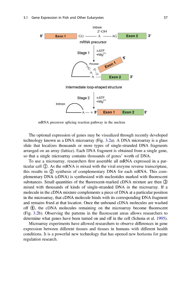
Fig. 3.1 mRNA precursor splicing reaction pathway in the nucleus
3.1 Gene Expression in Fish and Other Eukaryotes
57
technology known as a DNA microarray (Fig. 3.2a). A DNA microarray is a glass
slide that localizes thousands or more types of single-stranded DNA fragments
arranged on an array (lattice). Each DNA fragment is obtained from a single gene,
so that a single microarray contains thousands of genes’ worth of DNA.
To use a microarray, researchers first assemble all mRNA expressed in a particular cell ①. As the mRNA is mixed with the viral enzyme reverse transcriptase,
this results in ② synthesis of complementary DNA for each mRNA. This complementary DNA (cDNA) is synthesized with nucleotides marked with fluorescent
substances. Small quantities of the fluorescent-marked cDNA mixture are then ③
mixed with thousands of kinds of single-stranded DNA in the microarray. If a
molecule in the cDNA mixture complements a piece of DNA at a particular position
in the microarray, that cDNA molecule binds with its corresponding DNA fragment
and remains fixed at that location. Once the unbound cDNA molecules are washed
off ④, the cDNA molecules remaining on the microarray become fluorescent
(Fig. 3.2b). Observing the patterns in the fluorescent areas allows researchers to
determine what genes have been turned on and off in the cell (Schena et al. 1995).
Microarray experiments have allowed researchers to observe differences in gene
expression between different tissues and tissues in humans with different health
conditions. It is a powerful new technology that has opened new horizons for gene
regulation research.
Fig. 3.1 mRNA precursor splicing reaction pathway in the nucleus
3.1 Gene Expression in Fish and Other Eukaryotes
57