

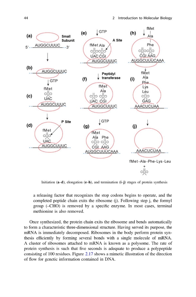
a releasing factor that recognizes the stop codons begins to operate, and the
completed peptide chain exits the ribosome (j). Following step j, the formyl
group (–CHO) is removed by a specific enzyme. In most cases, terminal
methionine is also removed.
Once synthesized, the protein chain exits the ribosome and bends automatically
to form a characteristic three-dimensional structure. Having served its purpose, the
mRNA is immediately decomposed. Ribosomes in the body perform protein synthesis efficiently by forming several bonds with a single molecule of mRNA.
A cluster of ribosomes attached to mRNA is known as a polysome. The rate of
protein synthesis is such that five seconds is adequate to produce a polypeptide
consisting of 100 residues. Figure 2.17 shows a mimetic illustration of the direction
of flow for genetic information contained in DNA.
Fig. 2.16 Initiation (a–d), elongation (e–h), and termination (i–j) stages of protein synthesis
44
2 Introduction to Molecular Biology
completed peptide chain exits the ribosome (j). Following step j, the formyl
group (–CHO) is removed by a specific enzyme. In most cases, terminal
methionine is also removed.
Once synthesized, the protein chain exits the ribosome and bends automatically
to form a characteristic three-dimensional structure. Having served its purpose, the
mRNA is immediately decomposed. Ribosomes in the body perform protein synthesis efficiently by forming several bonds with a single molecule of mRNA.
A cluster of ribosomes attached to mRNA is known as a polysome. The rate of
protein synthesis is such that five seconds is adequate to produce a polypeptide
consisting of 100 residues. Figure 2.17 shows a mimetic illustration of the direction
of flow for genetic information contained in DNA.
Fig. 2.16 Initiation (a–d), elongation (e–h), and termination (i–j) stages of protein synthesis
44
2 Introduction to Molecular Biology