

24 – Mollusca
281
(B)
(A)
(C)
(D)
(E)
(F)
(G)
(H)
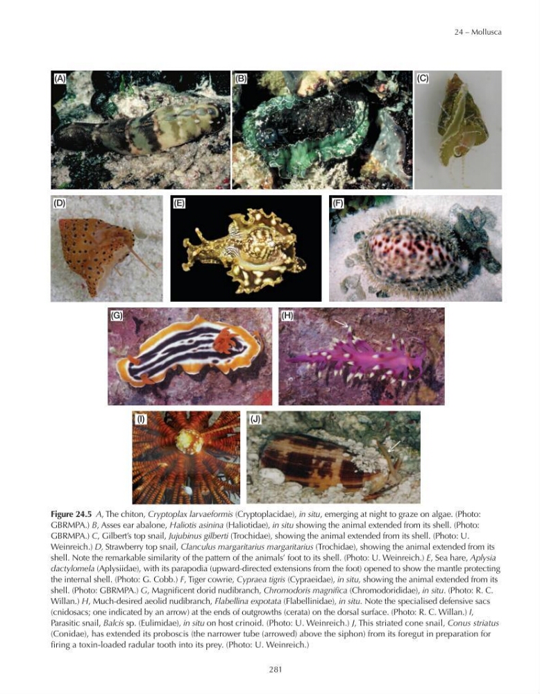
Figure 24.5 A, The chiton, Cryptoplax larvaeformis (Cryptoplacidae), in situ, emerging at night to graze on algae. (Photo:
GBRMPA.) B, Asses ear abalone, Haliotis asinina (Haliotidae), in situ showing the animal extended from its shell. (Photo:
GBRMPA.) C, Gilbert’s top snail, Jujubinus gilberti (Trochidae), showing the animal extended from its shell. (Photo: U.
Weinreich.) D, Strawberry top snail, Clanculus margaritarius margaritarius (Trochidae), showing the animal extended from its
shell. Note the remarkable similarity of the pattern of the animals’ foot to its shell. (Photo: U. Weinreich.) E, Sea hare, Aplysia
dactylomela (Aplysiidae), with its parapodia (upward-directed extensions from the foot) opened to show the mantle protecting
the internal shell. (Photo: G. Cobb.) F, Tiger cowrie, Cypraea tigris (Cypraeidae), in situ, showing the animal extended from its
shell. (Photo: GBRMPA.) G, Magnificent dorid nudibranch, Chromodoris magnifica (Chromodorididae), in situ. (Photo: R. C.
Willan.) H, Much-desired aeolid nudibranch, Flabellina expotata (Flabellinidae), in situ. Note the specialised defensive sacs
(cnidosacs; one indicated by an arrow) at the ends of outgrowths (cerata) on the dorsal surface. (Photo: R. C. Willan.) I,
Parasitic snail, Balcis sp. (Eulimidae), in situ on host crinoid. (Photo: U. Weinreich.) J, This striated cone snail, Conus striatus
(Conidae), has extended its proboscis (the narrower tube (arrowed) above the siphon) from its foregut in preparation for
firing a toxin-loaded radular tooth into its prey. (Photo: U. Weinreich.)
(I)
(J)
281
(B)
(A)
(C)
(D)
(E)
(F)
(G)
(H)
Figure 24.5 A, The chiton, Cryptoplax larvaeformis (Cryptoplacidae), in situ, emerging at night to graze on algae. (Photo:
GBRMPA.) B, Asses ear abalone, Haliotis asinina (Haliotidae), in situ showing the animal extended from its shell. (Photo:
GBRMPA.) C, Gilbert’s top snail, Jujubinus gilberti (Trochidae), showing the animal extended from its shell. (Photo: U.
Weinreich.) D, Strawberry top snail, Clanculus margaritarius margaritarius (Trochidae), showing the animal extended from its
shell. Note the remarkable similarity of the pattern of the animals’ foot to its shell. (Photo: U. Weinreich.) E, Sea hare, Aplysia
dactylomela (Aplysiidae), with its parapodia (upward-directed extensions from the foot) opened to show the mantle protecting
the internal shell. (Photo: G. Cobb.) F, Tiger cowrie, Cypraea tigris (Cypraeidae), in situ, showing the animal extended from its
shell. (Photo: GBRMPA.) G, Magnificent dorid nudibranch, Chromodoris magnifica (Chromodorididae), in situ. (Photo: R. C.
Willan.) H, Much-desired aeolid nudibranch, Flabellina expotata (Flabellinidae), in situ. Note the specialised defensive sacs
(cnidosacs; one indicated by an arrow) at the ends of outgrowths (cerata) on the dorsal surface. (Photo: R. C. Willan.) I,
Parasitic snail, Balcis sp. (Eulimidae), in situ on host crinoid. (Photo: U. Weinreich.) J, This striated cone snail, Conus striatus
(Conidae), has extended its proboscis (the narrower tube (arrowed) above the siphon) from its foregut in preparation for
firing a toxin-loaded radular tooth into its prey. (Photo: U. Weinreich.)
(I)
(J)