

ATPase, lipase, and cholinesterase. Inhibition of glycolysis, due in part to
decreased enolase activity, may be responsible for the hyperglycemia observed
in laboratory animals exposed to F.
F stimulates adenylcyclase activity in all tissues so far examined
(adenylcyclase catalyzes the formation of cyclic AMP [cAMP] from ATP). F
also affects functions controlled by Ca in humans, as it does in plants. These
functions include blood clotting, membrane permeability, and cholinesterase
activity. Fluoride inhibition of reactions involving Ca is generally attributed to
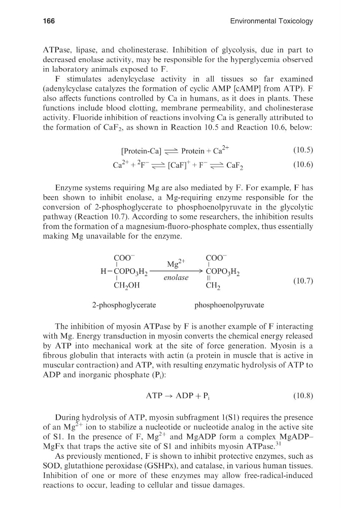
the formation of CaF 2 , as shown in Reaction 10.5 and Reaction 10.6, below:
ð10:5Þ
ð10:6Þ
Enzyme systems requiring Mg are also mediated by F. For example, F has
been shown to inhibit enolase, a Mg-requiring enzyme responsible for the
conversion of 2-phosphoglycerate to phosphoenolpyruvate in the glycolytic
pathway (Reaction 10.7). According to some researchers, the inhibition results
from the formation of a magnesium-fluoro-phosphate complex, thus essentially
making Mg unavailable for the enzyme.
ð10:7Þ
The inhibition of myosin ATPase by F is another example of F interacting
with Mg. Energy transduction in myosin converts the chemical energy released
by ATP into mechanical work at the site of force generation. Myosin is a
fibrous globulin that interacts with actin (a protein in muscle that is active in
muscular contraction) and ATP, with resulting enzymatic hydrolysis of ATP to
ADP and inorganic phosphate (P i ):
ATP ! ADP þ P i
ð10:8Þ
During hydrolysis of ATP, myosin subfragment 1(S1) requires the presence
of an Mg
2þ ion to stabilize a nucleotide or nucleotide analog in the active site
of S1. In the presence of F, Mg
2þ and MgADP form a complex MgADP–
MgFx that traps the active site of S1 and inhibits myosin ATPase.
31
As previously mentioned, F is shown to inhibit protective enzymes, such as
SOD, glutathione peroxidase (GSHPx), and catalase, in various human tissues.
Inhibition of one or more of these enzymes may allow free-radical-induced
reactions to occur, leading to cellular and tissue damages.
166
Environmental Toxicology
[16:52 26/8/04 P:/CRC PRESS/4365 MING-HO.751 (1670)/4365-010.3d]
Ref: 4365 MING-HO YU Chap-010 Page: 166 149-170
decreased enolase activity, may be responsible for the hyperglycemia observed
in laboratory animals exposed to F.
F stimulates adenylcyclase activity in all tissues so far examined
(adenylcyclase catalyzes the formation of cyclic AMP [cAMP] from ATP). F
also affects functions controlled by Ca in humans, as it does in plants. These
functions include blood clotting, membrane permeability, and cholinesterase
activity. Fluoride inhibition of reactions involving Ca is generally attributed to
the formation of CaF 2 , as shown in Reaction 10.5 and Reaction 10.6, below:
ð10:5Þ
ð10:6Þ
Enzyme systems requiring Mg are also mediated by F. For example, F has
been shown to inhibit enolase, a Mg-requiring enzyme responsible for the
conversion of 2-phosphoglycerate to phosphoenolpyruvate in the glycolytic
pathway (Reaction 10.7). According to some researchers, the inhibition results
from the formation of a magnesium-fluoro-phosphate complex, thus essentially
making Mg unavailable for the enzyme.
ð10:7Þ
The inhibition of myosin ATPase by F is another example of F interacting
with Mg. Energy transduction in myosin converts the chemical energy released
by ATP into mechanical work at the site of force generation. Myosin is a
fibrous globulin that interacts with actin (a protein in muscle that is active in
muscular contraction) and ATP, with resulting enzymatic hydrolysis of ATP to
ADP and inorganic phosphate (P i ):
ATP ! ADP þ P i
ð10:8Þ
During hydrolysis of ATP, myosin subfragment 1(S1) requires the presence
of an Mg
2þ ion to stabilize a nucleotide or nucleotide analog in the active site
of S1. In the presence of F, Mg
2þ and MgADP form a complex MgADP–
MgFx that traps the active site of S1 and inhibits myosin ATPase.
31
As previously mentioned, F is shown to inhibit protective enzymes, such as
SOD, glutathione peroxidase (GSHPx), and catalase, in various human tissues.
Inhibition of one or more of these enzymes may allow free-radical-induced
reactions to occur, leading to cellular and tissue damages.
166
Environmental Toxicology
[16:52 26/8/04 P:/CRC PRESS/4365 MING-HO.751 (1670)/4365-010.3d]
Ref: 4365 MING-HO YU Chap-010 Page: 166 149-170