

Relevant Methods for MRI/X Brain Image Registration
341
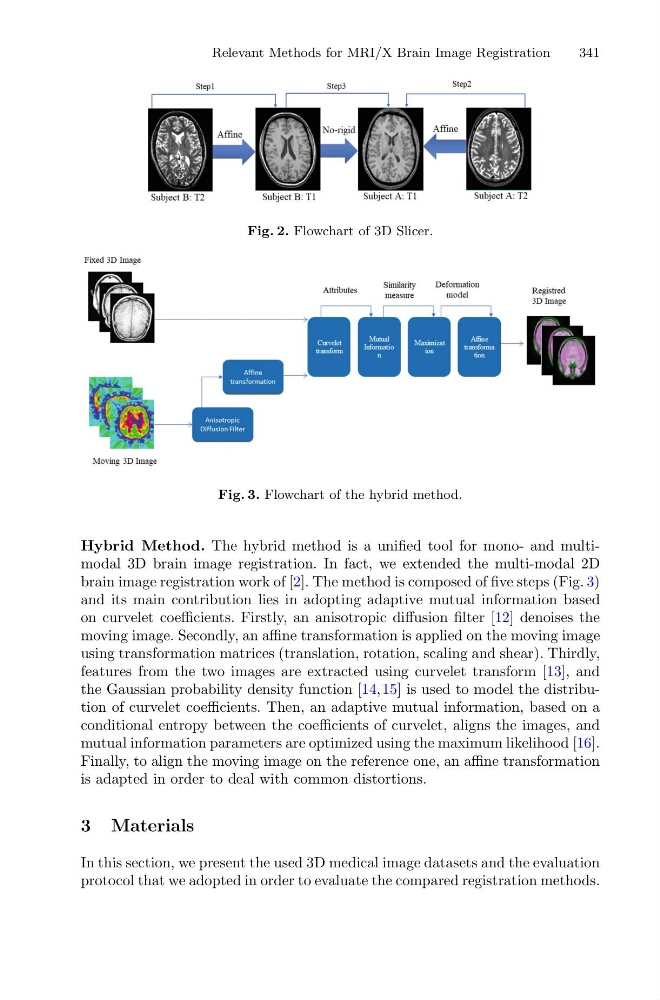
Fig. 2. Flowchart of 3D Slicer.
Fig. 3. Flowchart of the hybrid method.
Hybrid Method. The hybrid method is a unified tool for mono- and multimodal 3D brain image registration. In fact, we extended the multi-modal 2D
brain image registration work of [2]. The method is composed of five steps (Fig. 3)
and its main contribution lies in adopting adaptive mutual information based
on curvelet coefficients. Firstly, an anisotropic diffusion filter [12] denoises the
moving image. Secondly, an affine transformation is applied on the moving image
using transformation matrices (translation, rotation, scaling and shear). Thirdly,
features from the two images are extracted using curvelet transform [13], and
the Gaussian probability density function [14,15] is used to model the distribution of curvelet coefficients. Then, an adaptive mutual information, based on a
conditional entropy between the coefficients of curvelet, aligns the images, and
mutual information parameters are optimized using the maximum likelihood [16].
Finally, to align the moving image on the reference one, an affine transformation
is adapted in order to deal with common distortions.
3 Materials
In this section, we present the used 3D medical image datasets and the evaluation
protocol that we adopted in order to evaluate the compared registration methods.
341
Fig. 2. Flowchart of 3D Slicer.
Fig. 3. Flowchart of the hybrid method.
Hybrid Method. The hybrid method is a unified tool for mono- and multimodal 3D brain image registration. In fact, we extended the multi-modal 2D
brain image registration work of [2]. The method is composed of five steps (Fig. 3)
and its main contribution lies in adopting adaptive mutual information based
on curvelet coefficients. Firstly, an anisotropic diffusion filter [12] denoises the
moving image. Secondly, an affine transformation is applied on the moving image
using transformation matrices (translation, rotation, scaling and shear). Thirdly,
features from the two images are extracted using curvelet transform [13], and
the Gaussian probability density function [14,15] is used to model the distribution of curvelet coefficients. Then, an adaptive mutual information, based on a
conditional entropy between the coefficients of curvelet, aligns the images, and
mutual information parameters are optimized using the maximum likelihood [16].
Finally, to align the moving image on the reference one, an affine transformation
is adapted in order to deal with common distortions.
3 Materials
In this section, we present the used 3D medical image datasets and the evaluation
protocol that we adopted in order to evaluate the compared registration methods.