

340
M. Abderrahim et al.
)
b
(
)
a
(
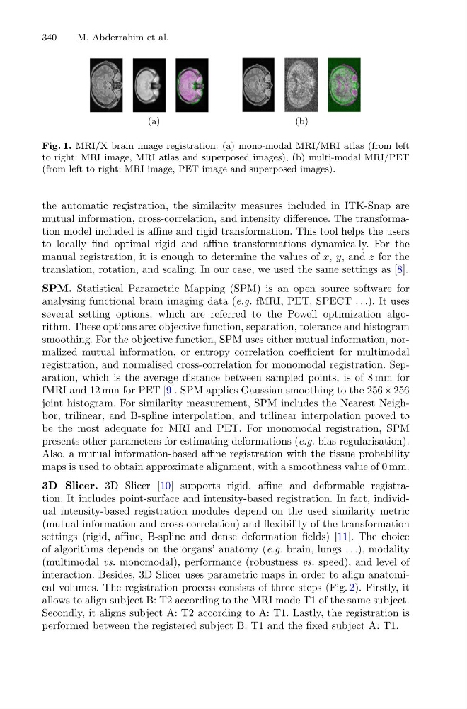
Fig. 1. MRI/X brain image registration: (a) mono-modal MRI/MRI atlas (from left
to right: MRI image, MRI atlas and superposed images), (b) multi-modal MRI/PET
(from left to right: MRI image, PET image and superposed images).
the automatic registration, the similarity measures included in ITK-Snap are
mutual information, cross-correlation, and intensity difference. The transformation model included is affine and rigid transformation. This tool helps the users
to locally find optimal rigid and affine transformations dynamically. For the
manual registration, it is enough to determine the values of x, y, and z for the
translation, rotation, and scaling. In our case, we used the same settings as [8].
SPM. Statistical Parametric Mapping (SPM) is an open source software for
analysing functional brain imaging data (e.g. fMRI, PET, SPECT . . .). It uses
several setting options, which are referred to the Powell optimization algorithm. These options are: objective function, separation, tolerance and histogram
smoothing. For the objective function, SPM uses either mutual information, normalized mutual information, or entropy correlation coefficient for multimodal
registration, and normalised cross-correlation for monomodal registration. Separation, which is the average distance between sampled points, is of 8 mm for
fMRI and 12 mm for PET [9]. SPM applies Gaussian smoothing to the 256 × 256
joint histogram. For similarity measurement, SPM includes the Nearest Neighbor, trilinear, and B-spline interpolation, and trilinear interpolation proved to
be the most adequate for MRI and PET. For monomodal registration, SPM
presents other parameters for estimating deformations (e.g. bias regularisation).
Also, a mutual information-based affine registration with the tissue probability
maps is used to obtain approximate alignment, with a smoothness value of 0 mm.
3D Slicer. 3D Slicer [10] supports rigid, affine and deformable registration. It includes point-surface and intensity-based registration. In fact, individual intensity-based registration modules depend on the used similarity metric
(mutual information and cross-correlation) and flexibility of the transformation
settings (rigid, affine, B-spline and dense deformation fields) [11]. The choice
of algorithms depends on the organs’ anatomy (e.g. brain, lungs . . .), modality
(multimodal vs. monomodal), performance (robustness vs. speed), and level of
interaction. Besides, 3D Slicer uses parametric maps in order to align anatomical volumes. The registration process consists of three steps (Fig. 2). Firstly, it
allows to align subject B: T2 according to the MRI mode T1 of the same subject.
Secondly, it aligns subject A: T2 according to A: T1. Lastly, the registration is
performed between the registered subject B: T1 and the fixed subject A: T1.
M. Abderrahim et al.
)
b
(
)
a
(
Fig. 1. MRI/X brain image registration: (a) mono-modal MRI/MRI atlas (from left
to right: MRI image, MRI atlas and superposed images), (b) multi-modal MRI/PET
(from left to right: MRI image, PET image and superposed images).
the automatic registration, the similarity measures included in ITK-Snap are
mutual information, cross-correlation, and intensity difference. The transformation model included is affine and rigid transformation. This tool helps the users
to locally find optimal rigid and affine transformations dynamically. For the
manual registration, it is enough to determine the values of x, y, and z for the
translation, rotation, and scaling. In our case, we used the same settings as [8].
SPM. Statistical Parametric Mapping (SPM) is an open source software for
analysing functional brain imaging data (e.g. fMRI, PET, SPECT . . .). It uses
several setting options, which are referred to the Powell optimization algorithm. These options are: objective function, separation, tolerance and histogram
smoothing. For the objective function, SPM uses either mutual information, normalized mutual information, or entropy correlation coefficient for multimodal
registration, and normalised cross-correlation for monomodal registration. Separation, which is the average distance between sampled points, is of 8 mm for
fMRI and 12 mm for PET [9]. SPM applies Gaussian smoothing to the 256 × 256
joint histogram. For similarity measurement, SPM includes the Nearest Neighbor, trilinear, and B-spline interpolation, and trilinear interpolation proved to
be the most adequate for MRI and PET. For monomodal registration, SPM
presents other parameters for estimating deformations (e.g. bias regularisation).
Also, a mutual information-based affine registration with the tissue probability
maps is used to obtain approximate alignment, with a smoothness value of 0 mm.
3D Slicer. 3D Slicer [10] supports rigid, affine and deformable registration. It includes point-surface and intensity-based registration. In fact, individual intensity-based registration modules depend on the used similarity metric
(mutual information and cross-correlation) and flexibility of the transformation
settings (rigid, affine, B-spline and dense deformation fields) [11]. The choice
of algorithms depends on the organs’ anatomy (e.g. brain, lungs . . .), modality
(multimodal vs. monomodal), performance (robustness vs. speed), and level of
interaction. Besides, 3D Slicer uses parametric maps in order to align anatomical volumes. The registration process consists of three steps (Fig. 2). Firstly, it
allows to align subject B: T2 according to the MRI mode T1 of the same subject.
Secondly, it aligns subject A: T2 according to A: T1. Lastly, the registration is
performed between the registered subject B: T1 and the fixed subject A: T1.