

90
S. Zorgui et al.
Until now, little works have been proposed for lentigo/healthy classification
of RCM images. In [10], the authors perform a two-dimensional wavelet decomposition. Then a generalized Gaussian distribution was applied to the wavelet
coefficients in order to perform a quantitative analysis assisted by a support
vector machine (SVM) to classify RCM images obtaining an accuracy of 84.4%.
Another approach in [11] explores a new unsupervised Bayesian algorithm for
the joint reconstruction and classification of RCM images. The resulting algorithm for healthy and lentigo classification reached an accuracy percentage of
97%. Beside, the paper [12] automatically diagnosed lentigo by using three separate feature extraction methods like Wavelets, Haralick and CNN by Transfer
Learning. The healthy/lentigo classification results reached an accuracy of 76%.
The present paper is organized as follows. Section 2 presents the problem
formulation of lentigo diagnosis. Section 3 detailed the proposed lentigo detection
method. Section 4 presents the experiment validation of our method. Finally,
conclusion and some perspectives are drawn in Sect. 5.
2 Related Work
2.1 Lentigo Detection
Lentigo is a lesion that occurs in the dermal epidermal junction between the
dermis and the epidermis involving a high concentration of melanocytes in the
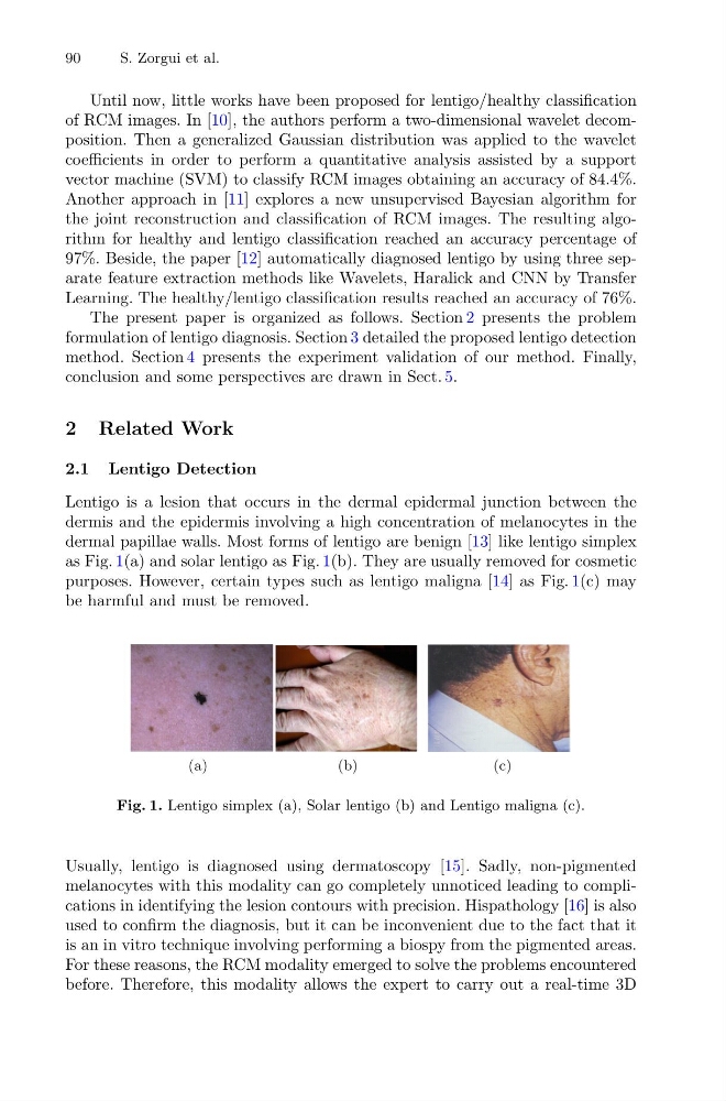
dermal papillae walls. Most forms of lentigo are benign [13] like lentigo simplex
as Fig. 1(a) and solar lentigo as Fig. 1(b). They are usually removed for cosmetic
purposes. However, certain types such as lentigo maligna [14] as Fig. 1(c) may
be harmful and must be removed.
(a)
(b)
(c)
Fig. 1. Lentigo simplex (a), Solar lentigo (b) and Lentigo maligna (c).
Usually, lentigo is diagnosed using dermatoscopy [15]. Sadly, non-pigmented
melanocytes with this modality can go completely unnoticed leading to complications in identifying the lesion contours with precision. Hispathology [16] is also
used to confirm the diagnosis, but it can be inconvenient due to the fact that it
is an in vitro technique involving performing a biospy from the pigmented areas.
For these reasons, the RCM modality emerged to solve the problems encountered
before. Therefore, this modality allows the expert to carry out a real-time 3D
S. Zorgui et al.
Until now, little works have been proposed for lentigo/healthy classification
of RCM images. In [10], the authors perform a two-dimensional wavelet decomposition. Then a generalized Gaussian distribution was applied to the wavelet
coefficients in order to perform a quantitative analysis assisted by a support
vector machine (SVM) to classify RCM images obtaining an accuracy of 84.4%.
Another approach in [11] explores a new unsupervised Bayesian algorithm for
the joint reconstruction and classification of RCM images. The resulting algorithm for healthy and lentigo classification reached an accuracy percentage of
97%. Beside, the paper [12] automatically diagnosed lentigo by using three separate feature extraction methods like Wavelets, Haralick and CNN by Transfer
Learning. The healthy/lentigo classification results reached an accuracy of 76%.
The present paper is organized as follows. Section 2 presents the problem
formulation of lentigo diagnosis. Section 3 detailed the proposed lentigo detection
method. Section 4 presents the experiment validation of our method. Finally,
conclusion and some perspectives are drawn in Sect. 5.
2 Related Work
2.1 Lentigo Detection
Lentigo is a lesion that occurs in the dermal epidermal junction between the
dermis and the epidermis involving a high concentration of melanocytes in the
dermal papillae walls. Most forms of lentigo are benign [13] like lentigo simplex
as Fig. 1(a) and solar lentigo as Fig. 1(b). They are usually removed for cosmetic
purposes. However, certain types such as lentigo maligna [14] as Fig. 1(c) may
be harmful and must be removed.
(a)
(b)
(c)
Fig. 1. Lentigo simplex (a), Solar lentigo (b) and Lentigo maligna (c).
Usually, lentigo is diagnosed using dermatoscopy [15]. Sadly, non-pigmented
melanocytes with this modality can go completely unnoticed leading to complications in identifying the lesion contours with precision. Hispathology [16] is also
used to confirm the diagnosis, but it can be inconvenient due to the fact that it
is an in vitro technique involving performing a biospy from the pigmented areas.
For these reasons, the RCM modality emerged to solve the problems encountered
before. Therefore, this modality allows the expert to carry out a real-time 3D