

240
9 Applications: Aqueous Interfaces
++
O
H
H
O
H
H
O
H
H
O
H
H
O
H
H
O
H H
Im[ ]
Im[ ]
SDS
CTAB
E z
E z
(2)
(2)
4
2
0
-2
-4
4
2
0
-2
-4
2800
3000
3200 3400
3600
2800
x10
-2
x10
-2
3000
3200 3400
3600
2 /cm -1
w
2 /cm -1
w
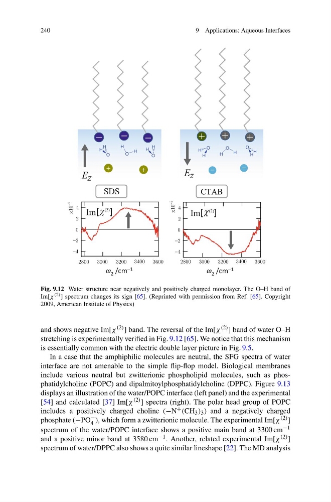
Fig. 9.12 Water structure near negatively and positively charged monolayer. The O–H band of
Im[χ (2) ] spectrum changes its sign [65]. (Reprinted with permission from Ref. [65]. Copyright
2009, American Institute of Physics)
and shows negative Im[χ (2) ] band. The reversal of the Im[χ (2) ] band of water O–H
stretching is experimentally verified in Fig. 9.12 [65]. We notice that this mechanism
is essentially common with the electric double layer picture in Fig. 9.5.
In a case that the amphiphilic molecules are neutral, the SFG spectra of water
interface are not amenable to the simple flip-flop model. Biological membranes
include various neutral but zwitterionic phospholipid molecules, such as phosphatidylcholine (POPC) and dipalmitoylphosphatidylcholine (DPPC). Figure 9.13
displays an illustration of the water/POPC interface (left panel) and the experimental
[54] and calculated [37] Im[χ (2) ] spectra (right). The polar head group of POPC
includes a positively charged choline (−N + (CH 3 ) 3 ) and a negatively charged
phosphate (−PO
−
4 ), which form a zwitterionic molecule. The experimental Im[χ (2) ]
spectrum of the water/POPC interface shows a positive main band at 3300 cm −1
and a positive minor band at 3580 cm −1 . Another, related experimental Im[χ (2) ]
spectrum of water/DPPC also shows a quite similar lineshape [22]. The MD analysis
9 Applications: Aqueous Interfaces
++
O
H
H
O
H
H
O
H
H
O
H
H
O
H
H
O
H H
Im[ ]
Im[ ]
SDS
CTAB
E z
E z
(2)
(2)
4
2
0
-2
-4
4
2
0
-2
-4
2800
3000
3200 3400
3600
2800
x10
-2
x10
-2
3000
3200 3400
3600
2 /cm -1
w
2 /cm -1
w
Fig. 9.12 Water structure near negatively and positively charged monolayer. The O–H band of
Im[χ (2) ] spectrum changes its sign [65]. (Reprinted with permission from Ref. [65]. Copyright
2009, American Institute of Physics)
and shows negative Im[χ (2) ] band. The reversal of the Im[χ (2) ] band of water O–H
stretching is experimentally verified in Fig. 9.12 [65]. We notice that this mechanism
is essentially common with the electric double layer picture in Fig. 9.5.
In a case that the amphiphilic molecules are neutral, the SFG spectra of water
interface are not amenable to the simple flip-flop model. Biological membranes
include various neutral but zwitterionic phospholipid molecules, such as phosphatidylcholine (POPC) and dipalmitoylphosphatidylcholine (DPPC). Figure 9.13
displays an illustration of the water/POPC interface (left panel) and the experimental
[54] and calculated [37] Im[χ (2) ] spectra (right). The polar head group of POPC
includes a positively charged choline (−N + (CH 3 ) 3 ) and a negatively charged
phosphate (−PO
−
4 ), which form a zwitterionic molecule. The experimental Im[χ (2) ]
spectrum of the water/POPC interface shows a positive main band at 3300 cm −1
and a positive minor band at 3580 cm −1 . Another, related experimental Im[χ (2) ]
spectrum of water/DPPC also shows a quite similar lineshape [22]. The MD analysis