

63
4 Raman optical Activity of Biological Samples
∆(
)
( )
[
( )
.
]
180
32
45
7
2
2
2
°
=
′
+
β
α
β α
G
c
(4.5b)
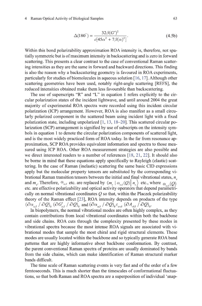
Within this bond polarizability approximation RoA intensity is, therefore, not spatially symmetric but is of maximum intensity in backscattering and is zero in forward
scattering. this presents a clear contrast to the case of conventional Raman scattering intensities as they are the same in forward and backward directions. this finding
is also the reason why a backscattering geometry is favoured in RoA experiments,
particularly for studies of biomolecules in aqueous solution [16, 17]. Although other
scattering geometries have been used, notably right-angle scattering [REFS], the
reduced intensities obtained make them less favourable than backscattering.
the use of superscripts “R” and “L” in equation 1 refers explicitly to the circular polarization states of the incident lightwave, and until around 2004 the great
majority of experimental RoA spectra were recorded using this incident circular
polarization (ICP) arrangement. however, RoA is also manifest as a small circularly polarized component in the scattered beam using incident light with a fixed
polarization state, including unpolarized [1, 13, 18–20]. this scattered circular polarization (SCP) arrangement is signified by use of subscripts on the intensity symbols in equation 1 to denote the circular polarization components of scattered light,
and is the most widely practiced form of RoA today. In the far from resonance approximation, SCP RoA provides equivalent information and spectra to those measured using ICP RoA. other RoA measurement strategies are also possible and
we direct interested readers to a number of references [18, 21, 22]. It should also
be borne in mind that these equations apply specifically to Rayleigh (elastic) scattering. In the case of Raman (inelastic) scattering the same basic CId expressions
apply but the molecular property tensors are substituted by the corresponding vibrational Raman transition tensors between the initial and final vibrational states, n v
and m v . therefore, α αβ etc. are replaced by 〈m
Q n
v
v
|
( )
α αβ
etc., where α αβ ( )
Q
etc. are effective polarizability and optical activity operators that depend parametrically on normal vibrational coordinates Q so that, within the Placzek polarizability
theory of the Raman effect [23], RoA intensity depends on products of the type
(
/
) (
/
)
α αβ
αβ
Q
G
Q
0
0 and
0
0
Q
A
Q
(
/
)
(
/
)
αβ
αγδ
γδβ
α
ε
.
In biopolymers, the normal vibrational modes are often highly complex, as they
contain contributions from local vibrational coordinates within both the backbone
and side chains. RoA cuts through the complexity presented by these modes in
vibrational spectra because the most intense RoA signals are associated with vibrational modes that sample the most chiral and rigid structural elements. these
modes are usually located within the backbone and so typically generate RoA band
patterns that are highly informative about backbone conformation. By contrast,
the parent conventional Raman spectra of proteins are usually dominated by bands
from the side chains, which can make identification of Raman structural marker
bands difficult.
the time scale of Raman scattering events is very fast and of the order of a few
femtoseconds. this is much shorter than the timescales of conformational fluctuations, so that both Raman and RoA spectra are a superposition of individual ‘snap-
4 Raman optical Activity of Biological Samples
∆(
)
( )
[
( )
.
]
180
32
45
7
2
2
2
°
=
′
+
β
α
β α
G
c
(4.5b)
Within this bond polarizability approximation RoA intensity is, therefore, not spatially symmetric but is of maximum intensity in backscattering and is zero in forward
scattering. this presents a clear contrast to the case of conventional Raman scattering intensities as they are the same in forward and backward directions. this finding
is also the reason why a backscattering geometry is favoured in RoA experiments,
particularly for studies of biomolecules in aqueous solution [16, 17]. Although other
scattering geometries have been used, notably right-angle scattering [REFS], the
reduced intensities obtained make them less favourable than backscattering.
the use of superscripts “R” and “L” in equation 1 refers explicitly to the circular polarization states of the incident lightwave, and until around 2004 the great
majority of experimental RoA spectra were recorded using this incident circular
polarization (ICP) arrangement. however, RoA is also manifest as a small circularly polarized component in the scattered beam using incident light with a fixed
polarization state, including unpolarized [1, 13, 18–20]. this scattered circular polarization (SCP) arrangement is signified by use of subscripts on the intensity symbols in equation 1 to denote the circular polarization components of scattered light,
and is the most widely practiced form of RoA today. In the far from resonance approximation, SCP RoA provides equivalent information and spectra to those measured using ICP RoA. other RoA measurement strategies are also possible and
we direct interested readers to a number of references [18, 21, 22]. It should also
be borne in mind that these equations apply specifically to Rayleigh (elastic) scattering. In the case of Raman (inelastic) scattering the same basic CId expressions
apply but the molecular property tensors are substituted by the corresponding vibrational Raman transition tensors between the initial and final vibrational states, n v
and m v . therefore, α αβ etc. are replaced by 〈m
Q n
v
v
|
( )
α αβ
etc., where α αβ ( )
Q
etc. are effective polarizability and optical activity operators that depend parametrically on normal vibrational coordinates Q so that, within the Placzek polarizability
theory of the Raman effect [23], RoA intensity depends on products of the type
(
/
) (
/
)
α αβ
αβ
Q
G
Q
0
0 and
0
0
Q
A
Q
(
/
)
(
/
)
αβ
αγδ
γδβ
α
ε
.
In biopolymers, the normal vibrational modes are often highly complex, as they
contain contributions from local vibrational coordinates within both the backbone
and side chains. RoA cuts through the complexity presented by these modes in
vibrational spectra because the most intense RoA signals are associated with vibrational modes that sample the most chiral and rigid structural elements. these
modes are usually located within the backbone and so typically generate RoA band
patterns that are highly informative about backbone conformation. By contrast,
the parent conventional Raman spectra of proteins are usually dominated by bands
from the side chains, which can make identification of Raman structural marker
bands difficult.
the time scale of Raman scattering events is very fast and of the order of a few
femtoseconds. this is much shorter than the timescales of conformational fluctuations, so that both Raman and RoA spectra are a superposition of individual ‘snap-