

T.P. Wróbel et al.
512
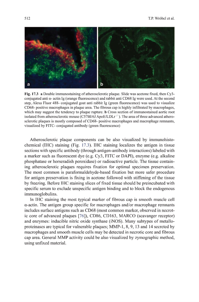
Atherosclerotic plaque components can be also visualized by immunohistochemical (IhC) staining (Fig. 17.3). IhC staining localizes the antigen in tissue
sections with specific antibody (through antigen-antibody interactions) labeled with
a marker such as fluorescent dye (e.g. Cy3, FItC or dAPI), enzyme (e.g. alkaline
phosphatase or horseradish peroxidase) or radioactive particle. the tissue containing atherosclerotic plaques requires fixation for optimal specimen preservation.
the most common is paraformaldehyde-based fixation but more safer procedure
for antigen preservation is fixing in acetone followed with stiffening of the tissue
by freezing. Before IhC staining slices of fixed tissue should be preincubated with
specific serum to exclude unspecific antigen binding and to block the endogenous
immunoglobulins.
In IhC staining the most typical marker of fibrous cap is smooth muscle cell
α-actin. The antigen group specific for macrophages and/or macrophage remnants
includes surface antigens such as Cd68 (most common marker, observed in necrotic core of advanced plaques [76]), Cd86, Cd163, mARCo (scavanger receptor)
and enzymes: inducible nitric oxide synthase (iNoS). many subtypes of metalloproteinases are typical for vulnerable plaques; mmP-1, 8, 9, 13 and 14 secreted by
macrophages and smooth muscle cells may be detected in necrotic core and fibrous
cap area. general mmP activity could be also visualized by zymographic method,
using unfixed material.
Fig. 17.3 a double immunostaining of atherosclerotic plaque. Slide was acetone fixed, then Cy3conjugated anti α- actin Ig (orange fluorescence) and rabbit anti CD68 Ig were used. At the second
step, Alexa Fluor 488- conjugated goat anti rabbit Ig (green fluorescence) was used to visualize
Cd68- positive macrophages in plaque area. the fibrous cap is highly infiltrated by macrophages,
which may suggest the tendency to plaque rupture. b Cross section of immunostained aortic root
isolated from atherosclerotic mouse (C57Bl/6J ApoE/LdLr
−/−
). the area of three advanced atherosclerotic plaques is mostly composed of Cd68- positive macrophages and macrophage remnants,
visualized by FItC- conjugated antibody (green fluorescence)
512
Atherosclerotic plaque components can be also visualized by immunohistochemical (IhC) staining (Fig. 17.3). IhC staining localizes the antigen in tissue
sections with specific antibody (through antigen-antibody interactions) labeled with
a marker such as fluorescent dye (e.g. Cy3, FItC or dAPI), enzyme (e.g. alkaline
phosphatase or horseradish peroxidase) or radioactive particle. the tissue containing atherosclerotic plaques requires fixation for optimal specimen preservation.
the most common is paraformaldehyde-based fixation but more safer procedure
for antigen preservation is fixing in acetone followed with stiffening of the tissue
by freezing. Before IhC staining slices of fixed tissue should be preincubated with
specific serum to exclude unspecific antigen binding and to block the endogenous
immunoglobulins.
In IhC staining the most typical marker of fibrous cap is smooth muscle cell
α-actin. The antigen group specific for macrophages and/or macrophage remnants
includes surface antigens such as Cd68 (most common marker, observed in necrotic core of advanced plaques [76]), Cd86, Cd163, mARCo (scavanger receptor)
and enzymes: inducible nitric oxide synthase (iNoS). many subtypes of metalloproteinases are typical for vulnerable plaques; mmP-1, 8, 9, 13 and 14 secreted by
macrophages and smooth muscle cells may be detected in necrotic core and fibrous
cap area. general mmP activity could be also visualized by zymographic method,
using unfixed material.
Fig. 17.3 a double immunostaining of atherosclerotic plaque. Slide was acetone fixed, then Cy3conjugated anti α- actin Ig (orange fluorescence) and rabbit anti CD68 Ig were used. At the second
step, Alexa Fluor 488- conjugated goat anti rabbit Ig (green fluorescence) was used to visualize
Cd68- positive macrophages in plaque area. the fibrous cap is highly infiltrated by macrophages,
which may suggest the tendency to plaque rupture. b Cross section of immunostained aortic root
isolated from atherosclerotic mouse (C57Bl/6J ApoE/LdLr
−/−
). the area of three advanced atherosclerotic plaques is mostly composed of Cd68- positive macrophages and macrophage remnants,
visualized by FItC- conjugated antibody (green fluorescence)