

T.P. Wróbel et al.
510
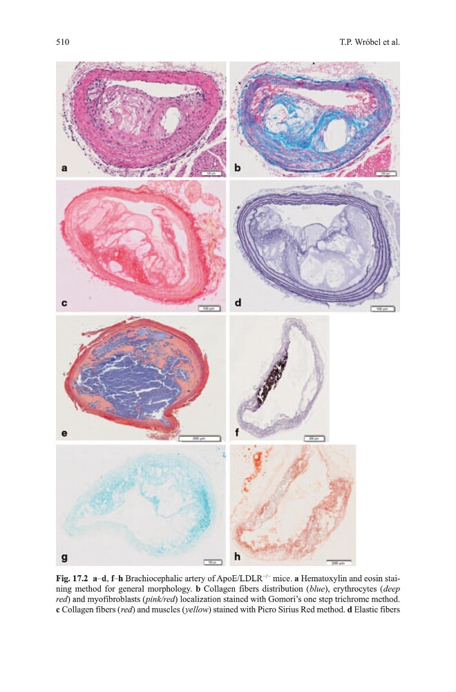
Fig. 17.2 a–d, f–h Brachiocephalic artery of ApoE/LdLR
−/−
mice. a hematoxylin and eosin staining method for general morphology. b Collagen fibers distribution ( blue), erythrocytes ( deep
red) and myofibroblasts ( pink/red) localization stained with gomori’s one step trichrome method.
c Collagen fibers ( red) and muscles ( yellow) stained with Picro Sirius Red method. d Elastic fibers
510
Fig. 17.2 a–d, f–h Brachiocephalic artery of ApoE/LdLR
−/−
mice. a hematoxylin and eosin staining method for general morphology. b Collagen fibers distribution ( blue), erythrocytes ( deep
red) and myofibroblasts ( pink/red) localization stained with gomori’s one step trichrome method.
c Collagen fibers ( red) and muscles ( yellow) stained with Picro Sirius Red method. d Elastic fibers