

497
16 Fourier Transform Infrared (Ft-Ir) Spectroscopic Imaging …
16.4.3 Cancer of the Colon
Ft-IR imaging has been used in several labs for diagnosing colon cancer by the
chemometric analysis of the absorption spectra [71–73] distinguishing between
healthy and malignant colon tissues and colon cancer and colitis [74]. histopathological analysis of colon tissue samples has been done to classify the cell types in
the colon tissues with high accuracy and specificity, in order to detect colon cancer
as well as other diseases of the colon such as ulcerative colitis or Crohn’s disease.
Previously, transmission IR imaging along with Baysian classification had been
used to classify the basic cell types like epithelial cells, goblet cells, loose connective tissue and blood cells [75]. Infrared images were then acquired employing the
AtR Ft-IR mode and transmission high-definition (74 ×) mode, both of which
showed much higher spatial resolution. the resulting images, as seen in, demonstrrate details that are not accessible at present. the intestinal crypts could be visualised much more clearly and even the small cells like lymphocytes and intracellular
mucin (which are potential diagnostic markers for cancer) could be classified accurately. this new imaging capability is likely to prove most crucial for colon tissue.
In prior applications, Ft-IR absorption spectra and algorithms were employed in
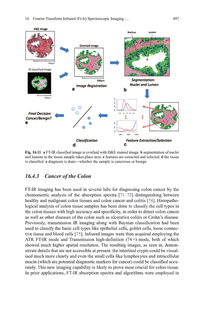
Fig. 16.11 a Ft-IR classified image is overlaid with h&E stained image. b segmentation of nuclei
and lumens in the tissue sample takes place next. c features are extracted and selected. d the tissue
is classified. e diagnosis is done—whether the sample is cancerous or benign
16 Fourier Transform Infrared (Ft-Ir) Spectroscopic Imaging …
16.4.3 Cancer of the Colon
Ft-IR imaging has been used in several labs for diagnosing colon cancer by the
chemometric analysis of the absorption spectra [71–73] distinguishing between
healthy and malignant colon tissues and colon cancer and colitis [74]. histopathological analysis of colon tissue samples has been done to classify the cell types in
the colon tissues with high accuracy and specificity, in order to detect colon cancer
as well as other diseases of the colon such as ulcerative colitis or Crohn’s disease.
Previously, transmission IR imaging along with Baysian classification had been
used to classify the basic cell types like epithelial cells, goblet cells, loose connective tissue and blood cells [75]. Infrared images were then acquired employing the
AtR Ft-IR mode and transmission high-definition (74 ×) mode, both of which
showed much higher spatial resolution. the resulting images, as seen in, demonstrrate details that are not accessible at present. the intestinal crypts could be visualised much more clearly and even the small cells like lymphocytes and intracellular
mucin (which are potential diagnostic markers for cancer) could be classified accurately. this new imaging capability is likely to prove most crucial for colon tissue.
In prior applications, Ft-IR absorption spectra and algorithms were employed in
Fig. 16.11 a Ft-IR classified image is overlaid with h&E stained image. b segmentation of nuclei
and lumens in the tissue sample takes place next. c features are extracted and selected. d the tissue
is classified. e diagnosis is done—whether the sample is cancerous or benign